
Open Access, Volume 11
Telomere length and disease-free longevity: Exploring the benefits of low-level laser therapy and nutritional support
Himanshu Bansal*; Mahima Mittal2; Anupama Bansal3; Mustafa Al Maini4; Matthias Wojcik5*; Robert Weber6; Martin Junggebauer6; Michael Weber6; Shahnawaz Hussein khan7
1Revita Lifesciences, Rudrapur 263153, India.
2Department of Functional Medicine, Lifeworth Hospital, Raipur 492001, India.
3HB Speciality Hospital & Research Institute, Rudrapur 263153, India.
4Department of Rheumatology, Mafraq Hospital, Abu Dhabi, United Arab Emirates.
5BSW Healthcare GmbH, 73079 Süßen, Germany.
6Weber Medical GmbH, 37697 Lauenförde, Germany.
7Consultant Neutrogennomic and Interventional Functional Medicine, Department of Regenerative Medicine, Lovinium Biotech,
Spain.
Himanshu Bansal
Revita Lifesciences, Rudrapur 263153, India.
Email: hbansal@drhbf.org
Received : October 13, 2025,
Accepted : November 24, 2025
Published : November 28, 2025,
Archived : www.jclinmedcasereports.com
Abstract
Background: Telomere shortening and cellular senescence are central features of biological aging and age-related decline. Although the human genome theoretically supports lifespans up to 120 years, most individuals do not reach this potential. Emerging evidence suggests that Low-Level Laser Therapy (LLLT), which mimics certain beneficial aspects of natural sunlight, can enhance telomere maintenance by stimulating cellular function and reducing oxidative stress.
Objective: To evaluate the effects of intravenous laser therapy combined with a targeted nutritional regimen on telomere length and regenerative biomarkers, with the goal of promoting disease-free lifespan extension.
Methodology: This prospective, open-label, single-arm intervention trial was conducted over three months in 30 healthy participants (aged 30-50 years). Baseline assessments included comprehensive health screenings and measurements of VSEL, CD34+, telomere length (TL), SDF-1, SCF, and VEGF. The same parameters were reassessed after three months. Data were analyzed using descriptive and inferential statistics.
Results: All measured parameters, VSEL levels (469.33±326.58 to 780.46±212.05), and CD34 levels (447.70±276.39 to 658.86±178.97), TL (9.71±0.86 to 10.31±0.93), SDF (1208.36±498.10 to 1608.66±423.40), SCF (1.01±0.29 to 1.25±0.24), and VEGF (84.15±33.09 to 119.76±20.59) showed significant improvements across all parameters. The Wilcoxon signed-rank test confirmed statistically significant improvements across all variables (p<0.001). These findings demonstrate a positive trend in the biological markers, suggesting potential implications for regenerative and therapeutic applications. Conclusion: The findings of this study give a preliminary indication that when IV laser application is integrated with the nutritional regimen, it is possible to improve the telomere length, augment the number of stem cells, and advance the favorable cytokine levels in 3 months. Collectively, these observations identify possible combined strategies for enhancing cellular vitality and reversing the aging process.
Keywords: VSEL cells; CD34+; Telomere length; VEGF; Regenerative biomarkers; Low-level laser therapy; Anti-aging interventions.
Copy right Statement: Content published in the journal follows Creative Commons Attribution License (http://creativecommons.org/licenses/by/4.0). © Bansal H (2025)
Journal: Open Journal of Clinical and Medical Case Reports is an international, open access, peer reviewed Journal mainly focused exclusively on the medical and clinical case reports.
Citation: Bansal H, Mittal M, Bansal A, Maini MA, Wojcik M, Weber R, Junggebauer M, et al. Telomere length and disease-free longevity: Exploring the benefits of low-level laser therapy and nutritional support. Open J Clin Med Case Rep. 2025; 2395.
Introduction
Human life expectancy has markedly increased due to medical advancements since the 19th century. Over the past two millennia, the average lifespan has extended, enhancing the likelihood that individuals may approach their lifespan potential. A crucial inquiry in this field is the maximum lifespan attainable by humans. The concept of longevity refers to living beyond the species-specific average age at death [1]. Historical evidence does not indicate an increase in the maximum human lifespan since recorded history began. The oldest recorded person, a woman who surpassed 122 years, often leads to the assumption that the maximum human lifespan is approximately 120 years [2]. Research by Dong suggests that the plausible maximum lifespan for humans may be around 115 years, emphasizing that this lifespan is likely fixed and constrained by biological factors. Moreover, investigations into optimal human lifespan indicate an inherent biological resistance to stress, positing that the realistic limits fall between 120 and 150 years [2]. Despite assertions that human lifespan is capped at around 122 years, other studies propose that there are no such fixed limits observed in animals [3].
The ongoing reduction in old-age mortality, coupled with an increase in the maximum age at death, may gradually enhance human longevity. Observations that lifespan in various animal species is flexible and can be extended through genetic or pharmaceutical interventions suggest that longevity might not be strictly constrained by species-specific genetics. In humans, the ends of chromosomes, known as telomeres, typically contain between 0.5 and 15 kilobases of identifiable repeating sequences [4], which are essential for maintaining genomic stability. Telomeres serve as protective caps at chromosome ends and shorten with each cell division; their length is considered a biomarker of biological aging. Research indicates that a healthy lifespan correlates positively with longer telomeres. Conversely, telomere dysfunction exacerbates the molecular hallmarks of aging [5]. Shorter lifespans have been linked to reduced telomere length [6], while the inverse relationship between age and telomere length in leukocytes positions telomeres as a cellular marker for biological aging (Muezzinler, Njajou). Various genetic, lifestyle, and environmental factors cumulatively contribute to the gradual decrease in telomere length over time, resulting in cellular senescence and the aging process [7].
Telomere loss is linked to an increased prevalence of chronic diseases, reduced cellular regenerative capacity, and heightened mortality rates. Schneider et al. (2022) highlighted a significant association between shorter telomere length [6] and elevated risks of cardiovascular diseases, cancer, and overall mortality, based on data from large cohort studies [8]. Furthermore, Adwan et al. (2019) proposed that telomeres may either contribute to longevity or result from it, emphasizing the bidirectional relationship between telomere biology and aging processes [9]. Research indicates that telomere attrition can be mitigated through various treatment methods, including ozone therapy, Hyperbaric Oxygen Therapy (HBOT), multivitamin supplementation, a balanced diet, and regular exercise, all of which may further delay aging. Interventions that enhance telomerase activity can potentially lengthen telomeres [10] and may aid in reversing or halting the aging process [11]. Additionally, nutritional status plays a crucial role in telomere maintenance, as numerous foods have been shown to influence TL through mechanisms related to oxidative stress, inflammation, DNA methylation, DNA integrity, and telomerase activity [12,13].
Long-term exposure to red and near-infrared light offers significant anti-inflammatory and therapeutic benefits, primarily through specific laser wavelengths that induce targeted cellular responses. Notably, Photobiomodulation (PBM) enhances mitochondrial function, reduces oxidative stress, and promotes cellular repair and regeneration, with laser treatment aiming to elicit these favorable cellular effects [14]. Current research focuses on utilizing these treatments to mitigate skin cell aging and reduce cellular stress. Additionally, chromophores such as melanin and hemoglobin interact with visible light, aiding in pigmentation and the regulation of inflammation [15]. However, while the effects of light and laser applications are becoming clearer, there is still limited concrete evidence demonstrating that PBM directly influences telomere length alteration, oxidative stress reduction, or cellular repair processes that could ultimately help maintain telomere length [16].
Genomic stability is essential for cell homeostasis, making it important to understand how laser application affects telomere elongation. Insights gained from this knowledge could enhance our understanding of intravenous laser therapy and nutritional interventions related to aging. In this study, we examine the impact of laser treatment and a nutritional regimen on telomere length as part of a life extension strategy. This research aims to establish a foundation for therapeutic targets based on telomere length, assuming that interventions designed to maintain or extend telomere length will yield health benefits.
Material and Methods
Study Design
This study was a prospective, open-label, single-arm intervention trial to evaluate the effects of laser application and nutritional regimen on telomere length for life extension.
Criteria: Healthy participants who voluntarily participated with no history of cancer or any previous telomere-targeted intervention were selected [10].
• The formula for age estimation: Age in years = 16.539 multiply mean TRF length + 236.287 [17].
• The relative telomere length RTL value was calculated as the ratio between the telomere signal of each sample and the control cell (TCL 1301 cell line) with correction for the DNA index of G0/1 cell [18].
Study protocol
Therapy is directed to the source of degeneration by repairing, regenerating, and restoring optimum function at the cellular level as well as the effective removal of the products of metabolism along with detoxification strategies. Our protocol can induce the expression of various growth factors, stem cell proliferation, mitochondrial biogenesis, angiogenesis and neurogenesis.
Patient assessment
- Comprehensive screening at baseline
- Complete evaluation of patient-specific issues.
- Evaluation based on family history based on risks.
- Hormone assays complete. Therapy Arm
Nutraceuticals and antioxidants
Patients were advised to take following nutraceutical combination for 12 weeks.
| Nutraceutical | Dose | Daily Dose |
|---|---|---|
| Acidophilus | 3 Billion | 1x |
| Alpha lipoic acid | 750 mg | 1x |
| Acetyl carnitine | 1000 mg | 1x |
| Alpha GPC | 600 mg | 1x |
| L-Arginine | 2000 mg | 1x |
| Aniracetam | 750 mg | 2x |
| Choline | 1000 mg | 1x |
| Coenzyme Q10 | 400 mg | 1x |
| DMAE | 200 mg | 3x |
| DHA | 2 g | 3x |
| EHA | 3 g | 1x |
| Ginko Biloba | 120 mg | 1x |
| N-Acetylcysteine | 600 mg | 4x |
| L-Taurine | 2 g | 1x |
| Vitamin C | 500 | 3x |
| Vitamin B Complex | 1 | 1x |
| Vitamin E | 5 | 3x |
| Curcumin | 500 mg | 2x |
Laser: IV Laser
IV Laser was applied for a total of 1h. Wavelengths/Colors were used for 5 days every week for 3 Months accordingly:
| Colours | Duration |
|---|---|
| Blue | 20 min |
| Green | 20 min |
| Yellow | 20 min |
| Red | 20 min |
| Infrared | 20 min |
Primary outcome measures
Vsel: Counts, E Cd34+ Counts, Telomeres Length Of Natural Killer cells, Levels Of Cytokines In Blood: Sdf-1 (Pg/Ml), Scf (Ng/Ml), Vegf (Pg/Ml), Sdf-1Stromal Cell Derived Factor and Scf Stem Cell Factor, Biochemical Analysis: Lipid Profile, Liver Function Test, Electrolytes, Blood Biochemistry, Hormone Levels and Hematology.
The quantity of Very Small Embryonic-Like stem cells (VSELs) in peripheral blood was determined, as these cells are believed to contribute to tissue repair and regeneration; their enumeration provides an estimate of the body’s regenerative potential [22]. Hematopoietic stem and progenitor cells (CD34+ cells) were also quantified to assess hematopoietic activity and the body’s capacity to generate new blood cells, with higher levels indicating enhanced regenerative competence and the potential for recovery from various disorders [23]. The telomere length of Natural Killer (NK) cells was measured as a biomarker of cellular aging and functional capacity, given that longer telomeres in NK cells are associated with improved immune surveillance and greater longevity, as observed in elderly cohorts [24]. Circulating cytokines, including Stromal cell-Derived Factor 1 (SDF-1), Stem Cell Factor (SCF), and Vascular Endothelial Growth Factor (VEGF), were analyzed and expressed in picograms per milliliter (pg/mL) or nanograms per milliliter (ng/mL). These cytokines play central roles in tissue repair, immune modulation, and intercellular communication [6,25]. In addition, comprehensive biochemical and safety assessments were performed, including lipid profiling, liver and kidney function tests, electrolyte panels, hormone analyses, and hematological evaluations [26,27]. Together, these parameters provided an integrated overview of organ function, metabolic status, and overall systemic health.
Secondary outcome measures
Attitude, Dfatigue, Sleep, Digestion and Appetite, Sexual Function and Frequency Questionnaires assessing attitudes explored participants’ psychological disposition and perceptions of health and aging, thereby capturing the influence of both positive and negative outlooks on overall health outcomes [28]. Fatigue was evaluated to quantify subjective experiences of tiredness or exhaustion, including potential contributions from underlying medical conditions associated with chronic fatigue [29]. Sleep quality was assessed using validated instruments, as restorative sleep is closely linked to physical well-being as well as cognitive and emotional functioning [30]. Digestive function and appetite were examined through structured questionnaires to identify gastrointestinal health status and potential nutritional deficiencies, both of which are relevant for systemic metabolic balance [31]. Finally, sexual function and frequency were evaluated, as these parameters are often affected by aging and provide valuable insights into overall vitality, quality of life, and general well-being [32].
Data collection and analysis
The data was collected from baseline and after 3 months of all variables. The data was entered and analyzed using SPSS Version 24. For continuous data, the descriptive are presented in mean±SD. The normality of data was assessed through Shapiro-Wilk and it was not normally distribution, therefore the Wilcoxon test was applied for within- group comparisons and the p-value≤0.05 was considered significant.
Results
The analysis included 30 participants, 17(56.7%) male and 13(43.3%) female, the mean age of participants was 45.3 years (SD=10.19), weight was 73.03 kg (SD=8.41), height was 5.49 feet (SD=0.26).
Table 1: Descriptive statistics of primary outcomes.
| Variables | N | Mean | Std. Deviation |
|---|---|---|---|
| VSEL Baseline | 30 | 469.33 | 326.58 |
| VSEL 3 Months | 30 | 780.46 | 212.05 |
| CD 34 Baseline | 30 | 447.70 | 276.39 |
| CD 34 3 Months | 30 | 658.86 | 178.97 |
| TL Baseline | 30 | 9.71 | 0.86 |
| TL 3 Months | 30 | 10.31 | 0.93 |
| SDF Baseline | 30 | 1208.36 | 498.10 |
| SDF 3 Months | 30 | 1608.66 | 423.40 |
| SCF Baseline | 30 | 1.01 | 0.29 |
| SCF 3 Months | 30 | 1.25 | 0.24 |
| VEGF Baseline | 30 | 84.15 | 33.09 |
| VEGF 3 Months | 30 | 119.76 | 20.59 |
| Valid N (listwise) | 30 |
(Table 1) presents baseline and 3-month follow-up measurements. VSEL levels increased from 469.33±326.58 at baseline to 780.46±212.05 after 3 months, while CD34 levels increased from 447.70±276.39 to 658.86±178.97. Telomere Length [19] also showed a positive change, from 9.71±0.86 to 10.31±0.93. Additionally, SDF levels increased from 1208.36±498.10 to 1608.66±423.40, SCF levels went up from 1.01±0.29 to 1.25±0.24, and VEGF levels shifted upward from 84.15±33.09 to 119.76±20.59. These results show a positive trend across all variables over the 3 months.
Table 2: Descriptive statistics of secondary outcomes.
| Statistics | |||||||
|---|---|---|---|---|---|---|---|
| Readings | Readings | Fatigue (FAS) | Sleep (SQS) | Digestion (DQLQ) | Sexual: (F Sfi): (lefi-5) | ||
| Baseline | N | Valid | 30 | 26 | 25 | 26 | 26 |
| Missing | 0 | 4 | 5 | 4 | 4 | ||
| Mean | 1.00 | 24.4231 | 33.6400 | 2.8077 | 17.4231 | ||
| Std. Deviation | .000 | 10.36985 | 17.00216 | .80096 | 7.56927 | ||
| Minimum | 1 | 10.00 | 4.00 | 2.00 | 2.00 | ||
| Maximum | 1 | 42.00 | 64.00 | 4.00 | 35.00 | ||
| 3 Months | N | Valid | 30 | 26 | 25 | 26 | 26 |
| Missing | 0 | 4 | 5 | 4 | 4 | ||
| Mean | 2.00 | 19.6923 | 26.2400 | 2.4615 | 22.9231 | ||
| Std. Deviation | .000 | 6.96143 | 10.99348 | .58177 | 4.04893 | ||
| Minimum | 2 | 9.00 | 9.00 | 2.00 | 11.00 | ||
| Maximum | 2 | 38.00 | 49.00 | 4.00 | 32.00 | ||
At baseline, the mean scores for fatigue (FAS), Sleep (SQS), Digestion (DQLQ), and sexual function (FSFI & IEFI-5) were 24.42, 33.64, 2.81, and 17.42, respectively. After three months, fatigue and sleep scores decreased to 19.69 and 26.24, indicating improvement, while sexual function scores increased to 22.92. This suggests a positive trend in overall well-being over the three months.
Table 3: Mean rank.
| Differences | N | Mean Rank | Sumof Ranks | Z | pvalues | |
|---|---|---|---|---|---|---|
| VSEL 3 Months – VSEL Baseline | Negative Ranks | 0 | 0 | 0 | -4.78 | 0 |
| Positive Ranks | 30 | 15.5 | 465 | |||
| Ties | 0 | |||||
| Total | 30 | |||||
| CD34 3 Months – CD 34 Baseline | Negative Ranks | 3 | 4 | 12 | -4.54 | 0 |
| Positive Ranks | 27 | 16.78 | 453 | |||
| Ties | 0 | |||||
| Total | 30 | |||||
| TL 3 Months – TL Baseline | Negative Ranks | 0 | 0 | 0 | -4.37 | 0 |
| Positive Ranks | 25 | 13 | 325 | |||
| Ties | 5 | |||||
| Total | 30 | |||||
| SDF 3 Months – SDF Baseline | Negative Ranks | 1 | 20 | 20 | -4.37 | 0 |
| Positive Ranks | 29 | 15.34 | 445 | |||
| Ties | 0 | |||||
| Total | 30 | |||||
| SCF 3 Months – SCF Baseline | Negative Ranks | 0 | 0 | 0 | -4.71 | 0 |
| Positive Ranks | 29 | 15 | 435 | |||
| Ties | 1 | |||||
| Total | 30 | |||||
| VEGF 3 Months – VEGF Baseline | Negative Ranks | 1 | 1 | 1 | -4.76 | 0 |
| Positive Ranks | 29 | 16 | 464 | |||
| Ties | 0 | |||||
| Total | 30 | |||||
(Table 3) shows the results of the Wilcoxon signed-rank test comparing the baseline and 3-month follow-up values. The test revealed statistically significant differences for all variables (VSEL, CD34, TL, SDF, SCF, VEGF), with Z values ranging from -4.371 to -4.782 and p-values of 0.000 for each, indicating that the 3-month follow-up values were significantly higher than baseline. The results confirm a significant improvement across all variables over the 3 months.
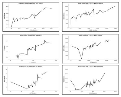
Figure 1: Graphical presentation of all variables (VSEL, CD34, TL, SDF, SCF, VEGF) comparing the baseline and 3-month follow-up values.
Para 1: Results of diabetes, liver, and thyroid conditions
Out of 30 study subjects, five were diabetic, three had hypothyroidism, and several exhibited liver dysfunction. Fasting sugar levels decreased from 139.2 mg/dL to 121.6 mg/dL, while post-meal glucose levels dropped from 259.6 mg/dL to 189 mg/dL. Among the diabetes patients, two (40%) experienced substantial decreases of over 25%, one (20%) had a moderate improvement of about 10%, and two (40%) showed minimal changes of less than 5%. In terms of thyroid function, TSH levels decreased from a mean of 6.1 mIU/L to 5.61 mIU/L, with two patients (66.7%) showing significant reductions of 8.33% and 13.43%, while one patient (33.3%) had a minimal change of 0.89%. Liver function tests indicated improvements as SGOT levels decreased from 77 U/L to 59.33 U/L and SGPT levels dropped from 79.33 U/L to 61.33 U/L. Two patients (66.7%) had significant reductions in SGOT of 33.33% and 30% for SGPT, while one (33.3%) showed minor reductions of 2.74% and 6.75%, respectively.
Para 2: VSEL and CD34 cell counts and growth factors
Significant increases were noted in both Very Small Embryonic-Like Stem Cells (VSELs) and CD34+ cell counts after three months of treatment. The VSEL count rose from a mean of 469.33/mL to 780.46/mL, representing a 66.3% increase. Among patients, one (3.3%) saw a minimal increase of less than 100/mL, eight (26.6%) showed increases between 400 and 600/mL, and the majority (70%) experienced increases of 300 to 400/mL. Similarly, CD34+ cell counts increased from 447.7/mL to 658.86/mL, indicating a 47.2% rise. Within this group, six (20%) recorded minimal increases below 100/mL, two (6.6%) had reductions, and approximately 70% saw increases between 100 and 300/mL. Over the same period, key growth factors such as SDF-1 increased by 33.1%, SCF rose by 23.9%, and VEGF saw a substantial improvement of 42.3%. These findings highlight an overall enhancement in signaling pathways and growth factor activity, contributing to improved cellular communication and tissue regeneration, alongside notable individual variability in responses.
Para 3: Questionnaire-based analysis of fatigue, sleep, digestion, and sexual function
In evaluating patient-reported outcomes, the mean Fatty Acid Synthase (FAS) levels showed a significant reduction, decreasing from an average of 24.42 to 19.69, equating to a 19.36% overall reduction. Three patients experienced declines of up to 50%. Sleep Quality Scores (SQS) improved significantly, dropping from a mean of 32.34 to 25.23, suggesting a 21.95% reduction in sleep disturbances. This indicates that the treatment effectively enhanced sleep quality for most participants. Additionally, scores for Digestion and appetite (DQLQ) displayed modest improvement, with the mean score decreasing from 2.80 to 2.46, reflecting a 12.14% reduction. This suggests positive advancements in digestive function and appetite regulation among the majority. Regarding sexual function and frequency, further analysis is needed to quantify improvements in this domain. Overall, these findings indicate a beneficial impact of the treatment across various health domains, improving both physiological measures and patient quality of life.
Discussion
Life expectancy, like other genetically determined features, varies from person to person since it is determined by genetics. It has been suggested that longevity may not be strictly subject to species-specific genetic constraints because the actual length of life is less than the potential life span, which reflects the influence of unfavorable environmental conditions. The study involved 30 participants with a mean age of 45.3 years, assessing the impact of interventions over three months. Significant increases were observed in various biomarkers critical to understanding cellular health and longevity. VSEL levels rose from 469.33 to 780.46, indicating enhanced levels of very small embryonic-like stem cells, which are thought to play a crucial role in tissue regeneration and repair.
Similarly, CD34 levels, which are markers for hematopoietic stem cells, increased from 447.70 to 658.86, reflecting improved cellular regeneration and a potentially more robust immune response. Telomere length, a vital indicator of cellular aging and genomic stability, improved from 9.71 to 10.31, suggesting that the interventions may aid in maintaining chromosomal integrity. Very Small Embryonic-Like Stem Cells (VSELs) (469.33 to 780.46) and CD34+ counts (447.70 to 658.86) increase fall in line with the observation that loss of stem cell number and activity over time drives organismal aging [33] it also corresponds to previous studies [34] which suggest that the intervention may enhance the body’s regenerative capacity. Additionally, this research demonstrated notable advancements in cytokines including SDF-1 (Stromal Cell-Derived Factor 1), SCF (Stem Cell Factor), and VEGF (Vascular Endothelial Growth Factor), suggesting improved cellular communication and angiogenesis.
SDF-1 is crucial for guiding stem cells and promoting tissue regeneration, while SCF is vital for the upkeep of hematopoietic stem cells [35]. Increased concentrations of SDF, SCF, and VEGF suggest enhanced support for angiogenesis and tissue mending, which may contribute to better overall health. Additionally, wellness assessments indicated notable progress in participant well-being, with fatigue scores decreasing from 24.42 to 19.69 and sleep quality scores increasing from 33.64 to 26.24. These changes suggest a reduction in overall stress levels and an improvement in restorative sleep, both essential for recovery and maintaining health. The rise in sexual function scores from 17.42 to 22.92 further indicates a boost in quality of life, likely linked to improvements in physical health and emotional wellness. The Wilcoxon signed-rank test validated significant statistical enhancements across all measured variables, with p-values at 0.000, confirming the effectiveness of the interventions. These results highlight the potential of targeted therapies to promote cellular vitality and enhance overall well-being in middle-aged adults.
The evaluation revealed significant improvements in health indicators among participants following the interventions. VSEL levels increased by 66.2%, while CD34 levels showed a 47.0% improvement, indicating enhanced regenerative capacity and a stronger immune response. Telomere length experienced a 6.2% improvement, suggesting better genomic stability. Additionally, SDF levels grew by 33.1%, SCF levels rose by 23.8%, and VEGF levels increased by 42.3%, pointing to enhanced support for angiogenesis and cellular healing processes. Participants reported a 19.0% reduction in fatigue and a 21.9% improvement in sleep quality. Furthermore, sexual function scores improved by 31.4%, indicating an overall enhancement in quality of life. These results demonstrate the significant effectiveness of the interventions, underscoring the substantial health benefits for participants over the three-month period.
In humans, significant evidence has been accumulated regarding the extension of lifespan through various methods, such as overnight fasting [36], calorie restriction [37,38], growth hormone supplementation [39], endurance training [40], weight loss, yoga, and stress management techniques, all of which have demonstrated meaningful correlations between antioxidant activity and telomerase activity but did not significantly alter telomere length [41-43]. Similarly, several studies have indicated that higher levels of physical activity or exercise are directly related to telomere length [44] and help maintain telomere length, with some research showing an increase in telomere length by as much as 5% [8]. Recently, a case report indicated that telomere length was doubled following HBO2 therapy, leading to a total increase of 100% in the telomere length of PBMCs. The telomere length of lymphocytes rose from 5.4 to 9 (a 66.7% increase), while the telomere length of monocytes grew from 5.8 to 12.7 (a 119.9% increase) [45]. Hadanny et al. also noted an average increase of 25-37% in lymphocyte telomere length among a group of 35 healthy elderly individuals [18].
Nutritional interventions, particularly the use of nutraceuticals, play a vital role in combating aging. Antioxidants like vitamins C and E, polyphenols, and dietary supplements such as resveratrol and curcumin can positively impact telomere dynamics by reducing oxidative stress and enhancing cellular repair mechanisms [46]. The length of the whole telomere genome was significantly increased (P<0.05) in experimental group (n=16 healthy volunteer) using special formulation containing Vit c natural vitamin E acetate 50%, vitamin B3, vitamin B12, β-carotene, vitamin B5, vitamin B6, vitamin B2, vitamin B1, folic acid, vitamin D3 biotin), Probiotics. 2,500 mg L-glutamine and 500 mg oligo fructose, 50 mg of ubiquinol, K2, magnesium. Omega 3-6-9, fish oil, linseed oil, borage oil Krill oil, alpha linolenic acid, eicosapentaenoic acid [5], docosahexaenoic acid [46], linolenic acid [47], linoleic acid and oleic acid [48].
Similarly, A two year trial conducted on cognitively healthy elderly adults, using a diet rich in walnuts, showed a non-significant trend to preserve telomere length when compared to a control diet [49]. Another study reported with omega-3 fatty acids, had an increased TL [47]. higher vitamin D concentrations, achieved with supplementation resulted in longer LTL [50]. It has been suggested that the mechanisms through which these nutritional factors attenuate telomere attrition are antioxidant activity, DNA methylation and the prevention of DNA damage [51]. In our study we corrected nutritional imbalance by covering all basic nutrients as for any regenerative effect nutrition has to be kept adequate. It could not be major bias S most of studies reported minimal effect. Nutritional supplements alone have reported benefits but small effects (2-5%) on TL [18].
Studies have already shown that the low-power laser could modify the expression of genes related to the nucleotide excision repair pathway, even in the skin and skeletal muscle of healthy Wistar rats, suggesting laser-induced modulation of DNA repair and genomic stability [52,53]. Such studies also corroborate results, suggesting that, depending on the laser parameters, the low-power laser irradiation can modulate the telomere length in healthy animals [54]. Laser therapy, particularly Low-Level Laser Therapy (LLLT), has attracted significant interest for its potential in reducing visible signs of aging, such as skin wrinkles, collagen degradation, and oxidative stress [55-58] however not yet studied in human for life extension despite lots of invitro supportive studies
Farias (2024) found that low-power therapeutic lasers and LEDs can alter telomere maintenance and length in human breast cancer cells [59]. Da Silva and collaborators (2023) showed the effects of low-power red laser and blue LED on three different cancer hallmarks at the same time [60]. The therapeutic mechanisms of LLLT are rooted in the absorption of light energy by mitochondrial cytochrome c oxidase, which catalyzes increased ATP production and bolstered cellular repair processes. However, LLLT has been studied for its effects in mitochondrial and cellular regeneration; its impact on telomere biology, however, remains not considered much. Mechanistically, LLLT may have a role in prevention of TL by reducing oxidative stress and inducing cellular repair. The beneficial effects of low-level laser application on the ARDS in animal models have been demonstrated [61-63] whereas this study results confirmed for humans. Effects of exposure to a low-power infrared laser on telomeres in heart tissue were extrapolated to humans [64], the low-power infrared laser irradiation increased the telomere length at 10 J cm−2 in cardiac tissue of animals affected by LPS- induced acute lung injury, and at delivering 3 J/cm2 to bone marrow mesenchymal stem cells which suggests that telomere maintenance is a part of the photobiomodulation effect induced by infrared radiation [54,65].
The evidence suggests that low-power laser exposure can alter telomere length, offering potential for life extension in healthy individuals. Combining IV laser treatment with dietary interventions may enhance cellular health by promoting tissue healing, reducing radical production, and boosting mitochondrial function [66]. Nutritional supplementation can address dietary deficiencies, support cellular metabolism, reduce inflammation, and prevent DNA damage. Together, these therapies may help prolong telomeres, increase stem cell numbers, and elevate protective cytokine levels [67]. This integrative approach targets multiple aging-related pathways, potentially leading to improved health outcomes and longevity, and highlights the importance of a holistic strategy that includes cellular repair, inflammation reduction, and nutritional support.
The integration of laser therapy’s ability to stimulate collagen production and reduce oxidative damage, alongside the antioxidant and anti-inflammatory properties of nutraceutical, presents a comprehensive approach to addressing the complexities of aging [68,69]. The synergy between Low-Level Laser Therapy (LLLT) and nutraceutical strategies has the potential to enhance longevity by targeting multiple pathways associated with telomere maintenance, oxidative stress reduction, inflammation, mitochondrial repair, and cellular regeneration [70].
This may represent a significant advancement in literature, providing a thorough evaluation of these therapeutic modalities, particularly within human models. However, despite promising preliminary results, telomere research continues to encounter several challenges. Most studies, such as those by Tsatsakis et al. (2023), are predominantly based on animal models, making it difficult to generalize findings to human populations [21]. Furthermore, existing trials have not adequately assessed long-term risks and benefits, as their durations remain relatively short. Schneider et al. (2022) emphasized the necessity for large-scale, longitudinal studies to establish telomere length as a reliable biomarker for disease prevention and the extension of health span [8].
Conducting comparative investigations of various anti-aging strategies is essential for pinpointing the most effective treatments. Although the current literature provides a broad understanding of how different interventions can improve both lifespan and health span, there is a pressing need for more comprehensive and systematically organized studies to yield conclusive results. Precision in methodology is crucial; differences in study design, participant demographics, and outcome assessments can significantly impact the results. Developing standardized criteria for evaluating aging, which includes measures of oxidative stress, skin elasticity, collagen production, and overall lifespan, will enable more accurate comparisons among various treatments. Research involving human subjects offers critical insights into the practical application and safety of these therapies. Conversely, studies conducted on animals provide valuable mechanistic insights and allow researchers to manipulate variables that may be difficult to control in human studies. Both research methods are extremely important, but there is an urgent need for integrated approaches that combine findings from both, to enhance our understanding of anti-aging mechanisms.
Conclusion
This prospective, open-label study provides preliminary evidence that the combined application of intravenous low-level laser therapy and a targeted nutritional regimen may beneficially modulate key biomarkers of cellular health and regeneration. Significant increases in VSEL and CD34+ stem cell counts, elongation of telomere length in natural killer cells, and elevated levels of regenerative cytokines (SDF-1, SCF, VEGF) suggest that the intervention has the potential to enhance regenerative capacity, support genomic stability, and improve systemic resilience. Importantly, these biological findings were paralleled by improvements in subjective health parameters, including reductions in fatigue, enhanced sleep quality, better digestive function, and improvements in sexual function. Collectively, these results highlight the potential of integrative strategies combining photobiomodulation and nutraceutical support to impact both objective biological markers and perceived quality of life.
From a translational perspective, the intervention appears safe and well tolerated, as demonstrated by stable biochemical, hormonal, and hematological parameters throughout the study period. This underscores the feasibility of implementing such protocols in broader clinical or wellness-oriented contexts. The observed improvements align with mechanistic insights from preclinical studies, supporting the hypothesis that low-level laser therapy, through mitochondrial stimulation and reduction of oxidative stress, may act synergistically with nutritional supplementation to slow cellular aging processes and promote tissue repair.
Nevertheless, several limitations must be acknowledged. The study design was single- arm and open-label, without a placebo or control group, making it difficult to exclude confounding factors or placebo-related effects. The sample size was relatively small (n=30) and restricted to healthy middle-aged adults, limiting generalizability to broader or clinical populations. The intervention period of three months, while sufficient to demonstrate measurable changes, does not address the sustainability of these effects over longer periods. Furthermore, lifestyle factors such as diet, physical activity, and genetic predispositions were not systematically controlled, which may have influenced individual responses.
Future research should therefore focus on larger, randomized controlled trials with longer follow-up durations to confirm the reproducibility and durability of these findings. Studies in patient populations with age-related diseases or compromised regenerative capacity will be particularly valuable to determine the therapeutic applicability of this combined intervention. In addition, mechanistic studies integrating multi-omics approaches could provide deeper insight into the molecular pathways through which intravenous laser therapy and nutritional supplementation interact to modulate telomere biology, stem cell dynamics, and systemic resilience.
In conclusion, this study offers encouraging preliminary data suggesting that integrative approaches targeting multiple hallmarks of aging may represent a promising avenue for extending disease-free lifespan. While the results are not yet sufficient to establish clinical efficacy, they provide a strong rationale for further, more rigorous investigation into the potential role of photobiomodulation combined with nutraceutical strategies in longevity and regenerative medicine.
Limitations and recommendations
The study design is single-arm, open-label making it impossible to separate the effects of the intervention from other factors. There could be some confounding factors or placebo effects could play a major part They have included only 30 participants and thus, the study lacks sufficient samples to produce the results on a large scale. The combination of the 3-month intervention and one-month follow-up may not detect the continuation of the changes in telomere length and other biomarkers. At the same time, vital valuables like diet, physical activity or genetic pre-dispositions to excessive fatness were not considered thus affecting the results. This evidence indicates that the use of RTL alone may fail to capture the general dynamics of aging sufficiently since it can only look at one parameter at a time. Future studies are suggested to conduct randomized controlled trials, longitudinal studies, comprehensive biomarker panels, personalized interventions, and mechanical studies to assess the long-term effects of an intervention on telomere length and stem cell populations, incorporating additional biomarkers for a holistic assessment.
References
- De Benedictis G, Franceschi C. The unusual genetics of human longevity. Sci Aging Knowledge Environ. 2006; 2006: pe20.
- Dong X, Milholland B, Vijg J. Evidence for a limit to human lifespan. Nature. 2016; 538: 257–259.
- Colchero F, Aburto J, Archie E, Boesch C, Breuer T, Campos F, et al. The long lives of primates and the ‘invariant rate of ageing’ hypothesis. Nat Commun. 2021; 12: 3666.
- Mumtaz S, Ali S, Tahir HM, Kazmi SAR, Shakir HA, Mughal TA, et al. Aging and its treatment with vitamin C: a comprehensive mechanistic review. Mol Biol Rep. 2021; 1–13.
- Chakravarti D, LaBella KA, DePinho RA. Telomeres: history, health, and hallmarks of aging. Cell. 2021; 184: 306–322.
- Kaszubowska L, Dettlaff-Pokora A, Hak L, Szarynska M, Ryba M, Mysliwska J, et al. Successful ageing of nonagenarians is related to the sensitivity of NK cells to activation. J Physiol Pharmacol. 2008; 59 Suppl 9: 187–199.
- Mason CE, Sierra MA, Feng HJ, Bailey SM. Telomeres and aging: on and off the planet. Biogerontology. 2024; 25: 313–327.
- Schneider CV, Schneider KM, Teumer A, Rudolph KL, Hartmann D, Rader DJ, et al. Association of telomere length with risk of disease and mortality. JAMA Intern Med. 2022; 182: 291–300.
- Adwan Shekhidem H, Sharvit L, Leman E, Manov I, Roichman A, Holtze S, et al. Telomeres and longevity: a cause or an effect. Int J Mol Sci. 2019; 20: 3233.
- Sharma A, Sane HM, Gokulchandran N, Kulkarni PP, Sayed H, Sawant D, et al. Anti-aging integrative therapy effects: a clinical study. 2024.
- Son DH, Park WJ, Lee YJ. Recent advances in anti-aging medicine. Korean J Fam Med. 2019; 40: 289.
- Galiè S, Canudas S, Muralidharan J, García-Gavilán J, Bulló M, Salas-Salvadó J. Impact of nutrition on telomere health: systematic review of observational cohort studies and randomized clinical trials. Adv Nutr. 2020; 11: 576–601.
- Tucker LA. Dietary fiber and telomere length in 5674 US adults: an NHANES study of biological aging. Nutrients. 2018; 10: 400.
- Randhawa M, Seo I, Liebel F, Southall MD, Kollias N, Ruvolo E. Visible light induces melanogenesis in human skin through a photoadaptive response. PLoS One. 2015; 10: e0130949.
- Tsibadze A, Chikvaidze E, Katsitadze A, Kvachadze I, Tskhvediani N, Chikviladze. Visible light and human skin. Georgian Med News. 2015; 246: 46–53.
- He X, Jin S, Dai X, Chen L, Xiang L, Zhang C. The emerging role of visible light in melanocyte biology and skin pigmentary disorders. J Clin Med. 2023; 12: 7488.
- Kumar V, Prakash A, Pandya P, Raina A. Telomere: a forensic approach for age estimation. Anil Aggrawal’s Internet J Forensic Med Toxicol. 2019; 20: 1.
- Hachmo Y, Hadanny A, Hamed RA, Daniel-Kotovsky M, Catalogna M, Fishlev G, et al. Hyperbaric oxygen therapy increases telomere length and decreases immunosenescence in isolated blood cells: a prospective trial. Aging (Albany NY). 2020; 12: 22445.
- Atli M, Engin-Ustun Y, Tokmak A, Caydere M, Hucumenoglu S, Topcuoglu C. Dose dependent effect of resveratrol in preventing cisplatin-induced ovarian damage in rats: an experimental study. Reprod Biol. 2017; 17: 274–280.
- Pallauf K, Rimbach G, Rupp PM, Chin D, Wolf IM. Resveratrol and lifespan in model organisms. Curr Med Chem. 2016; 23: 4639–4480.
- Tsatsakis A, Renieri E, Tsoukalas D, Buga AM, Sarandi E, Vakonaki E, et al. A novel nutraceutical formulation increases telomere length and activates telomerase activity in middle-aged rats. Mol Med Rep. 2023; 28: 1–11.
- Ratajczak M, Zuba-Surma E, Wojakowski W, Suszynska M, Mierzejewska K, Liu R, et al. Very small embryonic-like stem cells represent a real challenge in stem cell biology. Leukemia. 2014; 28: 473–484.
- Szilvassy SJ. The biology of hematopoietic stem cells. Arch Med Res. 2003; 34: 446–460.
- Bellantuono I. Haemopoietic stem cells. Int J Biochem Cell Biol. 2004; 36: 607–620.
- Pal J, Rajput Y, Shrivastava S, Gahine R, Mungutwar V, Barardiya T, et al. A standalone approach to utilize telomere length measurement as a surveillance tool in oral leukoplakia. Mol Oncol. 2022; 16: 1650–1660.
- Zachariah S, Kumar K, Lee SWH, Choon WY, Naeem S, Leong C. Interpretation of laboratory data and general physical examination by pharmacists. Clin Pharm Educ Pract Res. 2019; 91–108.
- Martens UM, Brass V, Engelhardt M, Glaser S, Waller CF, Lange W, et al. Measurement of telomere length in haematopoietic cells using in situ hybridization techniques. Biochem Soc Trans. 2000; 28: 245–250.
- Bryant C, Bei B, Gilson KM, Komiti A, Jackson H, Judd F. Antecedents of attitudes to aging: a study of the roles of personality and well-being. Gerontologist. 2016; 56: 256–265.
- Wessely S. Chronic fatigue: symptom and syndrome. Ann Intern Med. 2001; 134: 838–843.
- Gothe NP, Ehlers DK, Salerno EA, Fanning J, Kramer AF, McAuley E. Physical activity, sleep and quality of life in older adults. Behav Sleep Med. 2020; 18: 797–808.
- Romano C, Van Wynckel M, Hulst J, Broekaert I, Bronsky J, Dall’Oglio L, et al. ESPGHAN guidelines for gastrointestinal and nutritional complications in children with neurological impairment. J Pediatr Gastroenterol Nutr. 2017; 65: 242–264.
- Results from the Health Professionals Follow-up Study. Ann Intern Med. 2003;139:161–168.
- Schultz MB, Sinclair DA. When stem cells grow old: phenotypes and mechanisms of stem cell aging. Development. 2016;143:3–14.
- Baur JA, Sinclair DA. Therapeutic potential of resveratrol: the in vivo evidence. Nat Rev Drug Discov. 2006;5:493–506.
- Caiado F, Pietras EM, Manz MG. Inflammation as a regulator of hematopoietic stem cell function in disease, aging, and clonal selection. J Exp Med. 2021;218:e20201541.
- Helfand SL, de Cabo R. Evidence that overnight fasting could extend healthy lifespan. Nature. 2021;1–5.
- Hursting SD, Lavigne JA, Berrigan D, Perkins SN, Barrett JC. Calorie restriction, aging, and cancer prevention: mechanisms of action and applicability to humans. Annu Rev Med. 2003;54:131–152.
- Weyer C, Walford RL, Harper IT, Milner M, MacCallum T, Tataranni PA, et al. Energy metabolism after two years of energy restriction: the Biosphere 2 experiment. Am J Clin Nutr. 2000;72:946–953.
- Rudman D, Feller AG, Nagraj HS, Gergans GA, Lalitha PY, Goldberg AF, et al. Effects of human growth hormone in men over 60 years old. N Engl J Med. 1990;323:1–6.
- Werner CM, Hecksteden A, Morsch A, Zundler J, Wegmann M, Kratzsch J, et al. Differential effects of endurance, interval, and resistance training on telomerase activity and telomere length in a randomized, controlled study. Eur Heart J. 2019;40:34–46.
- Sanft T, Usiskin I, Harrigan M, Cartmel B, Lu L, Li FY, et al. Randomized controlled trial of weight loss versus usual care on telomere length in women with breast cancer. Breast Cancer Res Treat. 2018;172:105–112.
- Mason C, Risques RA, Xiao L, Duggan CR, Imayama I, Campbell KL, et al. Independent and combined effects of dietary weight loss and exercise on leukocyte telomere length in postmenopausal women. Obesity. 2013;21:E549–E554.
- Krishna BH, Keerthi GS, Kumar CK, Reddy NM. Association of leukocyte telomere length with oxidative stress in yoga practitioners. J Clin Diagn Res. 2015;9:CC01.
- Arsenis NC, You T, Ogawa EF, Tinsley GM, Zuo L. Physical activity and telomere length: impact of aging and potential mechanisms of action. Oncotarget. 2017;8:45008.
- Maroon JC. The effect of hyperbaric oxygen therapy on cognition, performance, proteomics, and telomere length. Front Neurol. 2022;13:949536.
- Epel ES, Blackburn EH, Lin J, Dhabhar FS, Adler NE, Morrow JD, et al. Accelerated telomere shortening in response to life stress. Proc Natl Acad Sci USA. 2004;101:17312–17315.
- Kiecolt-Glaser JK, Epel ES, Belury MA, Andridge R, Lin J, Glaser R, et al. Omega-3 fatty acids, oxidative stress, and leukocyte telomere length: a randomized controlled trial. Brain Behav Immun. 2013;28:16–24.
- Tsoukalas D, Fragkiadaki P, Docea AO, Alegakis AK, Sarandi E, Vakonaki E, et al. Association of nutraceutical supplements with longer telomere length. Int J Mol Med. 2019; 44: 218–226.
- Freitas-Simoes TM, Cofán M, Blasco MA, Soberón N, Foronda M, Serra-Mir M, et al. Walnut consumption for two years and leukocyte telomere attrition in Mediterranean elders. Nutrients. 2018; 10: 1907.
- Richards JB, Valdes AM, Gardner JP, Paximadas D, Kimura M, Nessa A, et al. Higher serum vitamin D concentrations are associated with longer leukocyte telomere length in women. Am J Clin Nutr. 2007; 86: 1420–1425.
- Herrmann M, Pusceddu I, März W, Herrmann W. Telomere biology and age-related diseases. Clin Chem Lab Med. 2018; 56: 1210–1222.
- de Souza da Fonseca A, Mencalha AL, Araújo de Campos VM, Ferreira Machado SC, de Freitas Peregrino AA, Geller M, et al. DNA repair gene expression in biological tissues exposed to low-intensity infrared laser. Lasers Med Sci. 2013; 28: 1077–1084.
- Scheller Madrid A, Rode L, Nordestgaard BG, Bojesen SE. Short telomere length and ischemic heart disease. Clin Chem. 2016; 62: 1140–1149.
- da Silva Neto Trajano LA, da Silva Sergio LP, de Oliveira DSL, Trajano ETL, dos Santos Silva MA, de Paoli F, et al. Low-power infrared laser modulates telomere length in heart tissue from an experimental model of acute lung injury. Photochem Photobiol Sci. 2021; 20: 653–661.
- Hernández-Bule ML, Naharro-Rodríguez J, Bacci S, Fernández-Guarino M. Unlocking the power of light on the skin: a comprehensive review on photobiomodulation. Int J Mol Sci. 2024; 25: 4483.
- Calderhead RG. Skin aging clock and its resetting by light-emitting diode low-level light therapy. 2017; 27.
- Beigvand HH, Razzaghi M, Rostami-Nejad M, Rezaei-Tavirani M, Safari S, Rezaei-Tavirani M, et al. Assessment of laser effects on skin rejuvenation. J Lasers Med Sci. 2020; 11: 212.
- Avci P, Gupta A, Sadasivam M, Vecchio D, Pam Z, Pam N, et al. Low-level laser therapy in skin: stimulating, healing, restoring. Semin Cutan Med Surg. 2013; 32: 41–52.
- Farias TG, Santos MS, Mencalha AL, da Fonseca AS. Low-power red laser and blue LED modulate telomere maintenance and length in human breast cancer cells. Lasers Med Sci. 2024; 39: 248.
- da Silva TG, Rodrigues JA, Siqueira PB, dos Santos Soares M, Mencalha AL, de Souza Fonseca A. Effects of photobiomodulation by low-power lasers and LEDs on the viability, migration, and invasion of breast cancer cells. Lasers Med Sci. 2023; 38: 191.
- Brown DL. Practical stereology applications for the pathologist. Veterinary Pathology. 2017; 54: 358–368.
- Martínez P, Gómez-López G, Pisano DG, Flores JM, Blasco MA. A genetic interaction between rap1 and telomerase reveals an unanticipated role for rap1 in telomere maintenance. Aging Cell. 2016; 15: 1113–1125.
- Oliveira Jr MC, Greiffo FR, Rigonato-Oliveira NC, Custódio RWA, Silva VR, Damaceno-Rodrigues NR, et al. Low level laser therapy reduces acute lung inflammation in a model of pulmonary and extrapulmonary LPS-induced ARDS. J Photochem Photobiol B. 2014; 134: 57–63.
- da Silva Neto Trajano LA, da Silva Sergio LP, de Oliveira DSL, Trajano ETL, dos Santos Silva MA, de Paoli F, et al. Low-power infrared laser modulates telomere length in heart tissue from an experimental model of acute lung injury. Photochem Photobiol Sci. 2021; 20: 653–661.
- Eroglu B, Genova E, Zhang Q, Su Y, Shi X, Isales C, et al. Photobiomodulation has rejuvenating effects on aged bone marrow mesenchymal stem cells. Sci Rep. 2021; 11: 13067.
- Nairuz T, Lee JH. Photobiomodulation therapy on brain: pioneering an innovative approach to revolutionize cognitive dynamics. Cells. 2024; 13: 966.
- Bjørklund G, Shanaida M, Lysiuk R, Butnariu M, Peana M, Sarac I, et al. Natural compounds and products from an anti-aging perspective. Molecules. 2022; 27: 7084.
- Ebrahiminaseri A, Sadeghizadeh M, Moshaii A, Asgaritarghi G, Safari Z. Combination treatment of dendrosomal nanocurcumin and low-level laser therapy develops proliferation and migration of mouse embryonic fibroblasts and alters TGF-β, VEGF, TNF-α and IL-6 expressions involved in wound healing process. PLoS One. 2021; 16: e0247098.
- Rossino MG, Casini G. Nutraceuticals for the treatment of diabetic retinopathy. Nutrients. 2019; 11: 771.
- Schellnegger M, Hofmann E, Carnieletto M, Kamolz LP. Unlocking longevity: the role of telomeres and its targeting interventions. Front Aging. 2024; 5: 1339317.
