
Open Access, Volume 11
Bowen’s disease in an unusual anatomical location: A case report
Neider Juliano Medina1; Jhon Anderson Zapata Vargas1; Luis Alejandro Sosa Ojeda1; Dayra Samira Morales Salcedo1; Mateo Andres Leal Capacho1; Ingrid Vanessa Ríos Anaya2; Miguel Ángel Durán Patiño3*
1General Medicine Practitioner, University of Pamplona, Norte de Santander, Colombia.
2Biologist, Universidad Industrial de Santander, Bucaramanga, Santander, Colombia.
3General Medicine Practitioner and Primary Care, Los Comuneros Hospital Universitario de Bucaramanga, Santander, University of Pamplona, Norte de Santander, Colombia.
Miguel Ángel Durán Patiño
General Medicine Practitioner and Primary Care, Los Comuneros University Hospital of Bucaramanga, Santander, Universidad de Pamplona, Norte de Santander, Colombia.
Email: mduran194@unab.edu.co
Received : October 01, 2025,
Accepted : November 04, 2025
Published : November 28, 2025,
Archived : www.jclinmedcasereports.com
Abstract
Introduction: Skin cancer is among the most common human neoplasms. It usually arises in areas exposed to ultraviolet radiation; however, its occurrence in non-exposed regions is associated with other etiological agents, such as HPV.
Clinical findings: An 83-year-old male presented with an 8-year history of a patch-like, flat lesion located on the external surface of the proximal third of the right thigh. The lesion measured approximately 3×3 cm, exhibited irregular borders, heterogeneous reddish-brown pigmentation, and mild scaling. Diagnosis, interventions, and outcomes: A diagnosis of Bowen’s disease was established. Wide local excision of the lesion was performed, resulting in complete remission.
Conclusion: Early detection of skin cancer in primary care, even in anatomically unusual locations, is essential to optimize clinical outcomes and prevent complications associated with Bowen’s disease.
Keywords: Skin cancer; Bowen’s disease; Older adult; HPV; Case report.
Abbreviations: HPV: Human Papilloma Virus; NMSC: Non-Melanoma Skin Cancers; BCC: Basal Cell Carcinoma; SCC: Squamous Cell Carcinoma; BD: Bowen’s Disease; UVR: Ultraviolet Radiation.
Copy right Statement: Content published in the journal follows Creative Commons Attribution License (http://creativecommons.org/licenses/by/4.0). © Patino MAD (2025)
Journal: Open Journal of Clinical and Medical Case Reports is an international, open access, peer reviewed Journal mainly focused exclusively on the medical and clinical case reports.
Citation: Medina NJ, Vargas JAZ, Ojeda LAS, Salcedo DSM, Capacho MAL, Anaya IVR, Patino MAD, et al. Bowen’s disease in an unusual anatomical location: A case report. Open J Clin Med Case Rep. 2025; 2391.
Introduction
Skin cancer is one of the most prevalent malignancies worldwide. It is traditionally classified according to histological features into NMSC, including basal BCC and SCC, and melanoma. SCC in situ is also referred to as BD. The primary risk factor is UVR exposure; nevertheless, genetic and infectious factors have also been implicated [1].
In 2022, a total of 19,976,499 new cancer cases were reported globally. Of these, 6.2% corresponded to NMSC and 1.7% to melanoma, with a male-to-female ratio of 3:2 [2]. In Colombia, during the same period, 58,813 new cases of all cancer types were documented, of which 19.4% were NMSC (14.7% BCC, 3.5% SCC, and 1.2% unspecified) and 1.1% melanoma. The overall mortality rates were 5.72 and 0.79 per 100,000 inhabitants, respectively, underscoring the significant public health burden of this condition [3].
SCC most commonly arises in sun-exposed areas such as the face and distal extremities, whereas BCC may develop in both photo-exposed and less exposed regions, such as the trunk. The thigh represents an uncommon anatomical site for both, and a rare site for BD [4]. Its proximity to the inguinal lymph node chain and the fact that it is infrequently examined in routine clinical practice increase the diagnostic challenge. Within this context, we report the case of an older adult with a large BD lesion located on the proximal third of the right thigh.
Case Presentation
An 83-year-old male, born and residing in Bucaramanga, unemployed, with a medical history of primary hypertension, primary hypothyroidism, and benign prostatic hyperplasia, presented in 2023 to his primary care physician with a cutaneous lesion described as a “spot” on the right thigh. The lesion had been present for 8 years, had enlarged over the past year, become pruritic, and changed in coloration. During this period, the patient had self-medicated with topical corticosteroids and clotrimazole without improvement.
On physical examination, a fair-skinned older adult was noted to have a flat, patch-like lesion measuring approximately 3×3 cm on the external surface of the proximal third of the right thigh. The lesion showed irregular borders, heterogeneous reddish-brown pigmentation, and mild scaling (Figure 1). No inguinal or popliteal lymphadenopathy was palpable, and no mucosal, genital, or additional cutaneous lesions were observed. He was referred to dermatology, where SCC was suspected, and a biopsy was performed. Histopathology confirmed SCC in situ (BD) with involvement of the lateral margins. The patient was subsequently referred to plastic surgery, where a wide local excision was carried out. The final pathology report demonstrated SCC in situ with clear margins. The patient remains under quarterly clinical follow-up with photoprotection. No metastases or perilesional lymphadenopathy were documented.
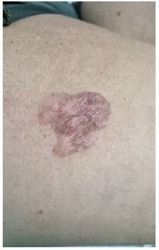
Figure 1: Photograph of the lesion. Bowen’s disease located on the lateral aspect of the proximal third of the right thigh. Original photograph.
Discussion
SCC in situ is characterized by disordered proliferation of keratinocytes confined to the epidermis, with BD representing one of its most frequent clinical variants. Although UV radiation is the principal risk factor, additional contributors include advanced age, HPV infection, immunosuppression, and certain medications, all of which may favor its occurrence in uncommon anatomical sites [5]. This case highlights such an unusual location, likely related to age and HPV infection, which, although not confirmed in this patient through laboratory testing, is known to be highly prevalent and underdiagnosed in males [6].
In primary care, the early recognition of cutaneous lesions such as BD is essential to prevent progression to invasive forms and to differentiate them from other skin cancers. The fact that this lesion remained undiagnosed for approximately eight years emphasizes the importance of timely diagnosis. Moreover, the lesion’s large size and proximity to a lymph node chain constituted significant risk factors for potential invasion and dissemination [7]. This underscores the need for thorough skin examinations and highlights the crucial role of comprehensive clinical evaluation in preventing serious complications. The patient’s self-medication with corticosteroids and antifungals, without success, further illustrates the lack of recognition of the lesion’s neoplastic nature, contributing to diagnostic delay.
The management of BD in this case consisted of wide surgical excision, an approach consistent with international guidelines [8].
Conclusion
This case underscores the importance of considering skin cancer in anatomically unusual locations. Early detection in primary care, combined with an appropriate multidisciplinary approach, is fundamental to improving clinical outcomes and preventing complications associated with Bowen’s disease.
Declarations
Acknowledgements: To the patient and his family.
References
- Wolff K, Johnson RA, Saavedra AP, Roh E. Fitzpatrick’s Color Atlas and Synopsis of Clinical Dermatology. 8th ed. New York, NY: McGraw-Hill Education LLC. 2017.
- International Agency for Research on Cancer. Cancer Today. Lyon: IARC; [cited 2024 Nov 13]. Available from: https://gco.iarc.who.int/today
- Fondo Colombiano de Enfermedades de Alto Costo (CAC). Situación del cáncer en la población adulta atendida en el SGSSS de Colombia, 2023. Bogotá, D.C.: CAC. 2024.
- Subramaniam P, Olsen CM, Thompson BS, Whiteman DC, Neale RE, for the QSkin Sun and Health Study Investigators. Anatomical Distributions of Basal Cell Carcinoma and Squamous Cell Carcinoma in a Population-Based Study in Queensland, Australia. JAMA Dermatol. 2017; 153: 175–82.
- Palaniappan V, Karthikeyan K. Bowen’s disease. Indian Dermatol Online J. 2022; 13: 177–89.
- Vives A, Cosentino M, Palou J. Evaluación del virus del papiloma humano en varones: primera revisión exhaustiva de la literatura. Actas Urol Esp. 2020; 44: 86–93.
- Caudill J, Thomas JE, Burkhart CG. The risk of metastases from squamous cell carcinoma of the skin. Int J Dermatol. 2023; 62: 483–6.
- Combalia A, Carrera C. Squamous Cell Carcinoma: An Update on Diagnosis and Treatment. Dermatol Pract Concept. 2020; 10: e2020066.
