
Open Access, Volume 9
Laboratory findings as milestones in crush syndrome
Mehmet Ali Berk Aridas1*; Ufuk Yukselmis2; Asuman Orcun1
1Department of Clinical Biochemistry, Kartal Dr. Lutfi Kırdar City Hospital, Istanbul, Türkiye.
2Department of Pediatric Intensive Care, Kartal Dr. Lutfi Kırdar City Hospital, Istanbul, Türkiye.
Mehmet Ali Berk Aridas
CDepartment of Clinical Biochemistry, Kartal Dr. Lutfi Kırdar City Hospital, Istanbul, Türkiye.
Tel: +905384893499;
Email: aliberkaridas97@gmail.com
Received : May 26, 2023,
Accepted : July 20, 2023
Published : July 25, 2023,
Archived : www.jclinmedcasereports.com
Abstract
We present a 9 years old case with complicated Crush Syndrome rescued from dent at 57th hour in Kahramanmaraş earthquake in Türkiye. We emphasized on the importance of laboratory support in all clinical stages of the patient; beginning from diagnosis through acute kidney injury and metabolic acidosis complications and follow-up of treatment in intensive care unit.
Keywords: Crush syndrome; Traumatic rhabdomyolysis; Acute kidney injury; Compartment syndrome.
Abbreviations: CS: Crush Syndrome; AKI: Acute Kidney Injury; PICU: Pediatric Intensive Care Unit.
Copy right Statement: Content published in the journal follows Creative Commons Attribution License (http://creativecommons.org/licenses/by/4.0). © Mehmet Ali BA (2023)
Journal: Open Journal of Clinical and Medical Case Reports is an international, open access, peer reviewed Journal mainly focused exclusively on the medical and clinical case reports.
Citation: Berk Aridas MA, Yukselmis U, Orcun A. Laboratory findings as milestones in crush syndrome. Open J Clin Med Case Rep. 2023; 2075.
Introduction
Crush Syndrome (CS) is a medical condition that includes rhabdomyolysis caused by trauma and associated surgical or medical signs and symptoms [1].
As a result of muscle trauma, destruction of the cell membrane leads to disruption of electrolyte regulation and the balance between the intracellular and extracellular milieu, release of constituents into the circulation and cell edema followed by cell lysis. When this process occurs throughout the muscle within a space bounded by fascia, compartment syndrome can result. Damage to cell membranes causes leaky capillary beds in compartment syndrome, which lowers intravascular volume in the injured limb. In the case of CS, sequestration of large volumes of fluid can lead to hypotension and hypovolemic shock. During cell lysis, the body faces a significant toxin load from the ischemic tissue, resulting in acidemia as well as electrolyte abnormalities, particularly hyperkalemia, hyperphosphatemia, and hypocalcemia. In addition to hypovolemia and rhabdomyolysis, renal injury is the third major component of CS. The etiology of renal injury is threefold. First, hypovolemia resulting from the previously described mechanisms causes renal hypoperfusion. Second, the release of myoglobin from damaged skeletal muscle in the course of rhabdomyolysis floods the plasma, supersaturating the haptoglobin molecules that normally bind it and increasing the filtration load of myoglobin on the renal tubules. Third, and most importantly, myoglobin itself is directly nephrotoxic [2].
The first cases of CS were reported during the 1909 Messina earthquake [3]. CS was first described after the Battle of London by Bywaters and Beall in 1941 [4]. CS typically occurs in war zones, mining disasters, after earthquakes, and in industrial and traffic accidents [5]. The incidence of CS has been reported to be 2% to 15% in all trauma patients and may be as high as 30% in earthquake victims [6]. The estimated proportion of patients requiring hemodialysis varies widely from 0% to 75% [2]. These rates, low at first glance, suggest that a large number of CS and related Acute Kidney Injury (AKI) cases may occur when the tens of thousands of post-disaster injuries are considered. For this reason, the laboratory parameters used in the treatment and monitoring of patients with CS are critical.
Case Presentation
Our case involves a 9-year-old male patient who was excavated from the dent at the 57th hour of the Kahramanmaraş-centered earthquake that occurred on February 06, 2023, in Türkiye. The patient was initially treated at Gaziantep State Hospital, fasciotomy was performed and necessary medical treatment was given, but the patient was referred to İstanbul Kartal Dr. Lütfi Kırdar City Hospital on February 9, 2023 due to the deepening of CS during follow-up examinations. He was directly interned in Pediatric Intensive Care Unit (PICU) because his general condition was poor and he was in a confused state. On the patient’s initial physical examination, he had compartment syndrome in the right foot, redness in the right thigh and ecchymosis of the left foot, left patella, and left heel. The abdomen had lacerations and ecchymosis of varying sizes. There was a fasciotomy scar in the lateral direction under the right patella and in the medial region of the right foot volar aspect. The patient’s right toes were cyanotic and the right foot and knee joint were immobile. On sensory examination, the right foot was numb, and the right Babinski reflex could not be detected.
The patient whose initial evaluation was completed was interned to the PICU. Due to CS induced-AKI, hemodialysis was planned for five days started from February 9, 2023. Amputation surgery was held on March 9, 2023 also received regular hyperbaric oxygen therapy. The patient, whose treatment and follow-up were conducted in PICU until March 13, 2023, then was transferred to the plastic and reconstructive surgery clinic for follow-up. On March 24, 2023, he was transferred to the orthopedic clinic for required physical therapy support.
Laboratory Evaluation
Laboratory test results are given in Table 1. Pathologic test results at the patient’s initial registration on February 9, 2023 were CK: 52126 U/L, LDH: 2011 U/L, AST: 1185 U/L, ALT: 367 U/L, urea: 149 mg/dL, creatinine: 1.69 mg/dL, uric acid: 8.8 mg/dL, calcium: 7.8 mg/dL, potassium: 6.08 mmol/ L, blood pH: 7.33, base excess: -4.0 mmol/L, HCO3: 20 mmol/L, lactate: 2.9 mmol/L, urine pH: 8.0, urine myoglobin: positive. As result of the patient’s effective treatments and the surgeries performed, the urine sample of March 1, 2023 showed a pH of 6.5 and a negative myoglobin value. In the blood gasses of March 13, 2023, pH: 7.41, HCO3: 24.9 mmol/L and lactate: 1.3 mmol/L were detected. In the blood sample of March 15, 2023, CK: 106 U/ L, LDH: 298 U/L, AST: 38 U/L, urea: 11 mg/dL, creatinine: 0.3 mg/dL, uric acid: 1.7 mg/dL, calcium: 8.7 mg/dL, potassium: 3.97 mmol/L were achieved.
It was observed that the regular fluid and hemodialysis treatments and amputation surgery performed in our case led to an improvement in both rhabdomyolysis and renal function tests. Monitorization of CK, LDH, AST, K, urea, and creatinine in serial blood samples drawn from our patient between February 09, 2023 and March 15, 2023 are shown in Figure 1. Muscle enzymes CK, AST and LDH related with rhabdomyolysis progressively decreased, reaching to normal levels parallel with their specific half lives. Urea, creatinine and K levels decreased as a result of effective hemodialysis treatment. Urine revealed myoglobin and urine alkalinization therapy to prevent heme and myoglobin deposition caused high urine pH. In the course of the circulatory support with fluid therapy, metabolic acidosis causing pH decrease and lactate increase was overcome.
Table 1: Laboratory parameters of the patient. First column presents the levels on the day of PICU admittance and second column represents the values on the day of PICU discharge.
| Tests | February 9, 2023 (Initial results) | March 13, 2023 (PICU discharge) | Referance Intervals |
|---|---|---|---|
| Blood pH | 7.33 | 7.41 | 7.35- 7.45 |
| Base Excess (mmol/L) | -4 | 3.1 | -1.5- +3 |
| HCO3 (mmol/L) | 20 | 24.9 | 22.5- 26.9 |
| Lactate (mmol/L) | 2.9 | 1.3 | 0.5-1.6 |
| CK (U/L) | 52126 | 119 | 0- 190 |
| LDH (U/L) | 2011 | 245 | 135- 225 |
| AST (U/L) | 1185 | 40 | 0- 40 |
| ALT (U/L) | 367 | 46 | 0- 41 |
| Urea (mg/dL) | 149 | 26 | 16.6- 48.5 |
| Creatinine (mg/dL) | 1.69 | 0.26 | 0.39- 0.73 |
| Urate (mg/dL) | 8.8 | 2.6 | 3.4- 7 |
| Calcium (mg/dL) | 7.8 | 8.9 | 8.8- 10.8 |
| Sodium (mmol/L) | 143 | 140 | 136- 145 |
| Potassium (mmol/L) | 6.08 | 4.01 | 3.5- 5.1 |
| Phosphorus (mg/dL) | 5.8 | 4.1 | 3.2- 5.7 |
| Magnesium (mg/dL) | 2.21 | 2.12 | 1.7- 2.1 |
| Chlorine (mmol/L) | 108 | 103 | 98- 107 |
| Urine myoglobin | +++ | Negative* | negative |
| Urine pH | 8.0 | 6.5* | 4.5 – 7.0 |
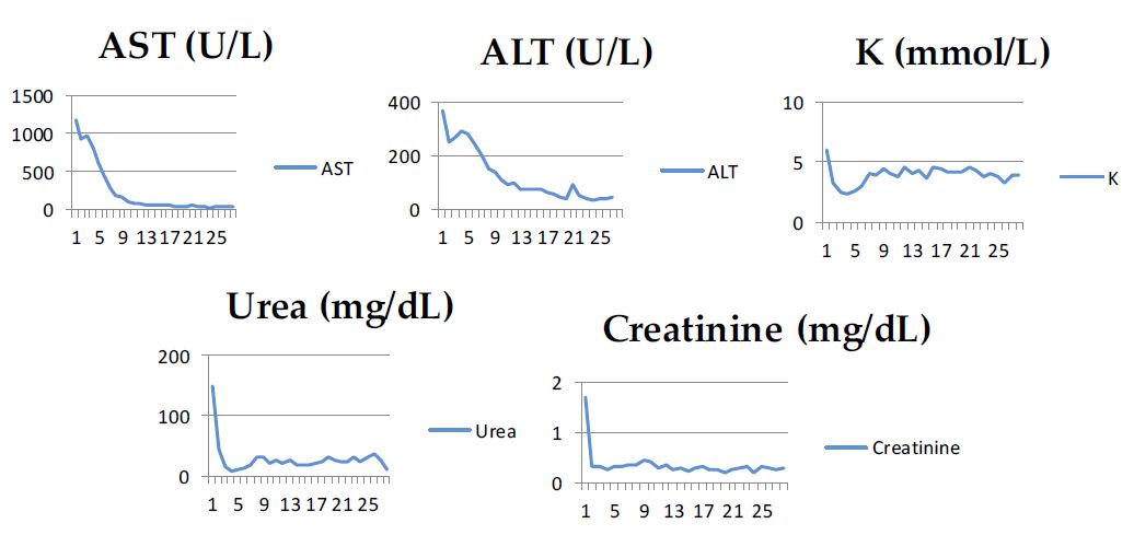
Figure 1: Serial monitorization of tests. X-axis show the 28 time points corresponding to days between February 9, 2023 and March 15, 2023. All of seven parameters were measured in each time point. Y-axis show the measured values of each test given in specific units presented in the figures.
Discussion
Laboratory evaluation of utmost importance at CS: Diagnosis is made with high levels of CK; more than 1000 U/L or above 5 times the upper limit of normal, along with urinary myoglobin. CK increases during injury and normalises rapidly. Its half-life is 48 hours. It is a reliable marker of muscle damage, but has no effect on other organs or the kidneys. Instead, myoglobin and heme are nephrotoxic and responsible for the progression of possible AKI [7].
Renal problems associated with CS have been defined as any of the following in association with oliguria/anuria; elevated levels of urea or creatinine, hyperkalemia, hyperphosphatemia, hypocalcemia, or metabolic acidosis [3].
Complications of rhabdomyolysis also include electrolyte imbalance, particularly hyperkalemia leading to cardiac arrhythmias, hyperphosphatemia, and hypocalcemia. Metabolic asidosis is the result of cell lysis leading to significant release of toxins from the ischemic tissue [7]. This is reflected in a pH decrease, HCO3 depletion, and negative base excess. Serum urate levels also increase as a result of impaired renal function.
Monitorization of these parameters during the clinical course is necessary and is a good measure of the patient's clinical condition and supports clinicians in treatment planning and patient management. Discharge criteria are based on clinical improvement, normal levels of electrolytes, creatine kinase, and creatinine levels, and normal urine output [8]. In cases with CS, more than one laboratory value contributes to patient management in this regard, beginning from diagnosis, through follow up of patient and finally discharge.
In the study by Dönmez et al. on CS children after the Marmara earthquake, Turkiye, 1999, laboratory parameters of 20 pediatric patients with CS were investigated and the efficiency and correlation of laboratory parameters such as CK, LDH, AST, ALT, phosphorus, creatinine, and D-dimer were evaluated. They found a positive correlation between CK and LDH, AST, ALT, phosphorus, creatinine and D-dimer [3]. In this study, the increase in serum K was also positively correlated with peak serum levels CK, AST, urea, and creatinine [3]. This suggested the severity of CS in these children. Similar results were found in the study by Oda et al after the 1995 Kobe earthquake [9]. In our case report, laboratory parameters were also well correlated with treatment and the clinical course of the patient.
Conclusion
CS is an important public health problem especially after earthquakes and consequences of rhabdomyolysis may be life threatening. Laboratory support in diagnosis,complications, treatment and follow up of this medical urgency is very important. Our case was particularly important because there is a limited number of literature on CS in pediatric age group.
Declarations
Acknowledgments: None to declare.
Funding: No funding source to disclose.
Declaration of conflicting interests: None declared.
References
- Sever L. Ezilme sendromu Çağrılı Editör. Türk Pediatri Arşivi. 2009; 44: 43-47.
- Lovallo E, Koyfman A, Foran M. Crush syndrome le syndrome de compression. African Journal of Emergency Medicine. 2012; 2: 117-123.
- Dönmez O, Meral A, Yavuz M, Durmaz O. Crush syndrome of children in the Marmara Earthquake, Turkey. Pediatr Int. 2001; 43: 678-682.
- Genthon A, Wilcox SR. Crush syndrome: A case report and review of the literature. J Emerg Med. 2014; 46: 313-319.
- Demirkiran O, Dikmen Y, Utku T, et al. Crush syndrome patients after the Marmara earthquakeEmergency Medicine Journal. 2003; 20: 247-250.
- Li W, Qian J, Liu X, et al. Management of severe crush injury in a front-line tent ICU after 2008 Wenchuan earthquake in China: an experience with 32 cases. Crit Care. 2009; 13: R178.
- Florou M, Lambropoulos V, Mouravas V, et al. Crush syndrome in a case of severe infant physical abuse: a case report. Pan Afr Med J. 2021; 39: 172.
- Szugye HS. Pediatric rhabdomyolysis. Pediatr Rev. 2020; 41: 265-275.
- Oda Y, Shindoh M, Yukioka H, Nishi S, Fujimori M, et al. Crush syndrome sustained in the 1995 Kobe, Japan, earthquake; treatment and outcome. Ann Emerg Med. 1997; 30: 507-512.
