
Open Access, Volume 9
Riga Fede Disease: A case report
Anusha Rudrakshi*; Naveen S; Praveenkumar Ramdurg; Surekha Puranik; EG Yog Naag Amaran; Saahil Ramesh
Department of Oral Medicine and Radiology, P M N M Dental College, and Hospital, Bagalkot, Karnataka, India.
Anusha Rudrakshi
Department of Oral medicine and Radiology, P M Nadagouda Memorial Dental College and Hospital,
S Nijalingappa Medical College and Hospital campus, Navanagar, Bagalkot, Karnataka-587103, India.
Email: anushabrudrakshi0@gmail.com
Received : May 15, 2023,
Accepted : July 07, 2023
Published : July 10, 2023,
Archived : www.jclinmedcasereports.com
Abstract
Riga-Fede disease is a condition in which the tongue moves back and forth over the mandibular anterior incisors due to the existence of natal-neonatal teeth or primary teeth, it frequently sustains traumatic damage. In the present case, a 5 month old female infant presented by parents to the department of Oral Medicine and Radiology with chief complaint of pain and a growth in the lower arch region of the mouth. Patient had occasional bleeding during breastfeeding for the past 15 days. Clinical examination revealed ulcerative overgrowth in the sublingual region measuring approximately 0.5 x 0.5 cm with indentation of the sharp incisal edge of the erupting 81 on the superior aspect of the growth. Based on history and clinical examination ‘Late RIGA- FEDE disease’ was given as provisional diagnosis. Treatment for Riga-Fede disease include conservative and surgical options and initially we opted for conservative treatment, but parents came back within 1 week with complaint of further discomfort in infant. Hence, extraction of 71 81 done on same day and recalled after 15 days for evaluation of ulcerative growth. We were able to determine at the follow-up that the lesion had fully regressed and the infant was feeding properly.
Keywords: Riga fede disease; Traumatic ulcer; Natal and neo-natal teeth; Infant and sublingual overgrowth.
Copy right Statement: Content published in the journal follows Creative Commons Attribution License (http://creativecommons.org/licenses/by/4.0). © Rudrakshi A (2023)
Journal: Open Journal of Clinical and Medical Case Reports is an international, open access, peer reviewed Journal mainly focused exclusively on the medical and clinical case reports.
Citation: Anusha R, Naveen S, Praveenkumar R, Surekha P, Naag Amaran EGY, et al. Riga Fede Disease: A Case Report. Open J Clin Med Case Rep. 2023; 2069.
Introduction
A rare benign mucosal lesion known as RIGA-FEDE disease is characterised by an ulcer on the ventral portion of the tongue brought on by frequent traumatic injuries due to tongue movement over the mandibular anterior teeth during breastfeeding [1].
Italian physician Antonio Riga first described the lesion in 1881; subsequently, F. Fede published the histological findings of the ulcer in 1890, which showed that it was an inflammatory lesion [2]. Since then, this condition has been known by a number of different names, including Riga’s disease, Riga-Fede disease or syndrome, sublingual granuloma, and traumatic sublingual ulceration [3]. A “PubMed” research revealed only 61 published cases of RFD in infants and children till now [4].
Riga-fede disease typically manifests as an ulcerative lesion on the ventral side of the tongue or in the sublingual region. It can also affect the lip, palate, gingiva, vestibular mucosa, and the floor of the mouth [5]. It can occasionally appear as sublingual growth in infants, lingual traumatic ulceration, traumatic atrophic glossitis, traumatic granuloma of the tongue, traumatic ulcerative granuloma with stromal eosinophilia, sublingual fibrogranuloma and fibrous hyperplasia on ventral surface of tongue [3].
RFD can be asymptomatic or sporadically linked with discomfort and bleeding from the lesion while feeding. RFD is most frequently linked to the eruption of the primary lower incisor in older infants or the natal-neonatal teeth in newborns [6]. It is uncommon for babies to have teeth at birth or within a month of delivery.
Occurrence of RFD is rare but the consequences make it an important condition for pediatricians and oral health professionals. Sublingual ulceration may resemble malignancies making it difficult to diagnosis. RFD also leads to multiple conditions like dehydration, malnutrition and painful nature of lesion is linked with incapacity to communicate, that can cause intellectual impairment [7]. Hence, timely diagnosis and treatment will prevent all the above mentioned complications.
Here, we are discussing about a case of RFD associated with early eruption of lower deciduous central incisors at the age of 4 months in a female infant and also discussing about its management.
Case Presentation
A five-month-old female infant reported to the Department of Oral Medicine and Radiology, P.M.N.M. Dental College and Hospital, Bagalkot, with a complaint of pain and a growth in the lower arch region of the mouth, with occasional bleeding during breastfeeding for the past 15 days, resulting in reduced food intake and deprived sleep in the recent 2-3 days.
On intraoral examination, a solitary ulcerative growth was seen in the sublingual region, situated exactly in the midline of the ventral part of tongue and corresponding to erupting 71 and 81. It measured approximately 0.5 cm X 0.5 cm and extends superioinferiorly from approximately 10 mm away from the tip of the elevated tongue to 5 mm away from the erupting 71 and 81. The superior aspect of the ulcerative growth showed an indentation from the sharp incisal edge of the erupting 81. On palpation, all inspection findings were confirmed with respect to site, size, and extent. The growth was soft to firm in consistency without any indurated margins, and an irregular border on the superior aspect and a smooth surface on the periphery. The infant showed slight discomfort and mild bleeding upon palpation. A sharp, irregular incisal edge was noted in relation to the erupting 81.
Based on clinical examination, Riga Fede disease was given as a provisional diagnosis, which presented as fibrous reactive overgrowth with superficial ulceration. A few differential diagnoses were considered, including infections like congenital syphilis, hematological disorders like agranulocytosis, and neoplasia such as myofibroma and vascular malformation [8].
In this case, initially conservative approach was followed by smoothing the incisal edge of tooth 81 and placing composite resin over the offending teeth. We advised the application of a local anesthetic gel containing benzocaine for 10 days. However, the parents returned within one week with complaints of further discomfort and bleeding from the same region. This resulted in reduced food intake and caused the infant to lose weight. Therefore, we performed the extraction of teeth 71 and 81 on the same day and advised the continuation of topical anesthetic for another 15 days. During the third visit, one month after the extraction, complete healing in the sublingual region of the oral cavity was noted. The parents of the treated child reported that infant was feeding normally and noticed improvement in systemic health within one month.
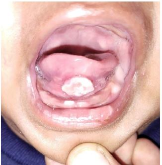
Figure 1: Traumatic ulcerative overgrowth in the sublingual region of the mouth corresponding to the erupting 71 and 81.

Figure 2: Complete regression of the lesion within 1 month of extraction of 71 and 81.
Discussion
The Riga-Fede Disease is a traumatic tongue ulceration brought on by recurrent trauma carried on by the tongue moving back and forth over the mandibular anterior incisors. Although ulcer can affect any surface of the oral mucosa, it is most commonly found in the ventral tongue region [1]. Failure to detect the lesions can result in medical complications such as deformity or tongue mutilation, insufficient nutritional intake, and dehydration, all of which can negatively affect growth and development [9].
There are two distinct categories for Riga-Fede syndrome. Before the age of six months, instances are considered to be "early" because they involve natal or neonatal teeth, which frequently have hypoplastic enamel and underdeveloped roots and thus erupt early. "Late" instances (those involving the primary dentition and occurring after six months of age) are frequently recurrent and may be connected to neurological or developmental conditions like familial dysautonomia [10] (Insensitivity to pain).
In our case, it is a "late Riga-Fede disease" that involved the early eruption of the mandibular primary central incisor at the age of 4 months, while the normal eruption sequence starts at 6 months. Most cases of Riga-Fede disease occur in infants with natal-neonatal teeth or with primary erupting teeth. Infants often
thrust their tongue over the alveolar ridge during feeding, resulting in frequent trauma from the sharp incisal edge of erupting teeth.
Due to frequent trauma, RFD usually begins as an ulcerated area with noticeable raised edges and can advance to an enlarged, fibrous mass that appears to be an ulcerative granuloma and has superficial necrosis. Histopathologically, RFD is distinguished by an ulcerated epithelium with granulation tissue and a mixed inflammatory infiltrate that includes mast cells, lymphocytes, macrophages, and a significant number of eosinophils. According to van der Meij et al. [8], once clinicians are familiar with RFD, the typical history and clinical features are usually sufficient, and additional histopathological examination is rarely necessary. In this case, we diagnosed relying solely on a complete history and physical examination while keeping the age of patient in mind and opted to treat conservatively first, if lesion is not subsided then we planned for extraction of offending tooth or excision of the lesion.
Infants with ulcerative growth of the oral mucosa may have a variety of differential diagnoses.
1. TRAUMATIC — mechanical (Riga Fede disease), electrical, chemical.
2. INFECTION — congenital syphilis, tuberculosis.
3. HEMATOLOGICAL DISORDER — agranulocytosis.
4. LOCAL NEOPLASIA — granular cell tumour, myofibroma, sarcoma, extra-nodal lymphoma, infantile hemangion, acute myeloid leukemia and other vascular malformation [8].
There are various treatments available for Riga Fede disease that aim to eradicate the cause of trauma in order to promote healing. It is recommended to initiate treatment with conservative approaches such as smoothing out the edges of the incisors, covering the rough edges with composite resin, modifying feeding patterns by using a bottle with a bigger hole in the nipple, placing a nasogastric tube, or administering a local corticosteroid to relieve symptoms are other options for treating rough edges [11]. The extraction of the incisors may be an option if these treatments fail to heal the lesion or if the child is seriously malnourished or dehydrated. Another option is excision of the lesion itself [11]. In this case, we opted for conservative treatment by smoothing out the incisal edge of tooth 81 and applying composite resin over the affected teeth and adviced for application of topical anesthetic for 10 days. Present case highlights the potential consequences of failing to diagnose as parents came back within a week with complaint of irritability and breastfeeding difficulties in an infant due to an injury on the ventral part of the tongue. Therefore, extraction of teeth 71 and 81 was done on the same day, and they were advised to continue using topical anesthetic for another 15 days. During the third visit, which was one month after the extraction, complete healing in the sublingual area of the oral cavity was noted, and infant was reported as healthy and active by the parents.
Conclusion
Traumatic ulcers can develop in the oral cavity, particularly in sublingual region of infants aged below 2 years. These ulcers may be caused by sharp edges of erupting teeth, including natal-neonatal or primary teeth. When the child thrusts their tongue on the mandibular anterior region during feeding, it can result in an ulcerative lesion or depending on the duration may be associated with fibrous overgrowth. Infants with prematurely erupted teeth should therefore undergo thorough examination to enable early diagnosis and appropriate treatment. Differential diagnosis plays an important role here because sometime lesion mimics the neoplastic feature which changes the complete treatment plan. Hence, we should rule out all possibilities of different systemic disease featuring same character as presented case. Additionally, parents should be counseled to raise awareness about the potential consequences of the presence of natal-neonatal teeth and the lesions that may cause health issues in their child.
References
- Costacurta M, Maturo P, Docimo R. Riga-Fede disease and neonatal teeth. Oral Implantol (Rome). 2012; 5: 26-30.
- Jamani NA, Ardini YD, Harun NA. Neonatal tooth with Riga-Fide disease affecting breastfeeding: A case report. Int Breastfeed J. 2018; 13: 35.
- Jingarwar MM, Bajwa NK, Pathak A. Riga Fede Disease: Fibrous Hyperplasia Associated with Natal Teeth in an Infant-A Case Report and Clinical Update. 2014; 2: 11-13.
- Mansur AT, Deniz K, Ozdemir K. ‘Riga-Fede disease like ulcers in old age: A case report/Ileri yasta Riga-Fede benzeri ulserler: Bir olgu sunumu’, Turkish Archives of Dermatology and Venereology. 2019; 53: 157.
- Biradar, Nandini. Rigafede and Neonatal teeth. International Journal of Medical Reviews and Case Reports. 2020; 5: 5455.
- Patil Sonal, Dixit Uma. A Trio Discovery-Cardarelli-Riga-Fede Disease: A Case Report and Review of Literature. Journal of Contemporary Dentistry. 2013; 3: 44-48.
- Volpato LE, Simoes CA, Simoes F, Nespolo PA, Borges AH. Riga-Fede Disease Associated with Natal Teeth: Two Different Approaches in the Same Case. Case reports in dentistry. 2015; 234961.
- van der Meij EH, de Vries TW, Eggink HF, de Visscher JGAM. Traumatic lingual ulceration in a newborn: Riga-Fede disease. Ital J Pediatr. 2012; 38.
- Mohan R P S, Verma S, Gill N, Singh U. Riga-Fede disease (Cardarelli’s aphthae): A report of nine cases. S. Afr. j. child health. 2014; 8: 72-74.
- Alahmari A, Albatool S. Management of Riga-Fede disease: A Case Report. Dentistry. 2017; 07.
- Baghdadi ZD. Riga-Fede disease: Association with microcephaly. International Journal of Paediatric Dentistry. 2002; 12: 442-445.
