
Open Access, Volume 9
A rare case of bullous leukocytoclastic vasculitis mimicking bullous pemphigoid
Aayush Pradhan; Roxana Oriana Florica*; Tamandeep Kaur; Anirudh Jaglan
Department of Western Reserve Health Education/Northeast Ohio Medical School (NEOMED), 1350 East Market Street, Warren, Ohio, 44483, USA.
Roxana Oriana Florica
Department of Western Reserve Health Education/Northeast Ohio Medical School (NEOMED), 1350 East Market Street, Warren, Ohio, 44483, USA.
Email: Roxana.florica@steward.org
Received : May 10, 2023,
Accepted : June 28, 2023
Published : June 30, 2023,
Archived : www.jclinmedcasereports.com
Abstract
Leukocytoclastic vasculitis is a small vessel vasculitis which typically presents with a painful, burning rash predominantly involving the lower extremities. The most common skin manifestation is palpable purpura. We present a case of a 61-year-old male who presented with tense bullae in bilateral lower extremities who was diagnosed with bullous leukocytoclastic vasculitis - one of the rare presentations of leukocytoclastic vasculitis. It should be differentiated from Bullous Pemphigoid which has a similar clinical manifestation using skin biopsy pathology. Definite treatment and prognosis of idiopathic bullous leukocytoclastic vasculitis remains largely unknown.
Keywords: Leukocytoclastic vasculitis; Small vessel vasculitis; Bullous pemphigoid; Palpable purpura; Fibrinoid necrosis.
Copy right Statement: Content published in the journal follows Creative Commons Attribution License (http://creativecommons.org/licenses/by/4.0). © Florica RO (2023)
Journal: Open Journal of Clinical and Medical Case Reports is an international, open access, peer reviewed Journal mainly focused exclusively on the medical and clinical case reports.
Citation: Pradhan A, Florica RO, Kaur T, Jaglan A. A rare case of bullous leukocytoclastic vasculitis mimicking bullous pemphigoid. Open J Clin Med Case Rep. 2023; 2064.
Introduction
Leukocytoclastic Vasculitis (LCV) refers to a small vessel vasculitis which typically involves capillaries, postcapillary venules, and arterioles with inflammatory infiltrates composed mainly of neutrophils with fibrinoid necrosis and degradation fragments of nucleic components. It predominantly affects the skin. The most commonly associated systemic diseases when LCV is suspected, are pANCA and cANCA associated vasculitides, connective tissue diseases, cryoglobuilinemic vasculitis, and Henoch-Schonlein purpura. Thus, an extensive work-up is imperative at the beginning of this clinical presentation to determine if it is a skin-limited or a skin-predominant presentation of another systemic disease.
Skin presentations in LCV typically appear as palpable purpura of lower extremities, however urticarial wheals, erythematous plaques, livedoid ulcerations and hemorrhagic vesiculobullous lesions are other less common manifestations. Several courses of disease evolution have been described. An acute appearance of simultaneous vasculitis lesions of the same age is often associated with an infection or drug-induced cause. Recurrent purpura with periods of complete resolution of symptoms can be seen in connective tissue diseases. Lastly, chronic persistent lesions of LCV are typically noted in patients with cryoglobulinemia, systemic small-vessel vasculitis, or malignancies.
In this case, we discuss a 61-year-old-male who presented with progressively increasing number of painful scattered palpable purpura and bullae of the lower extremities. It was initially suspected to be bullous pemphigoid, however skin punch biopsy revealed findings consistent with LCV. Our case is a rare manifestation of LCV-bullous type. Improvement of symptoms with systemic steroids was noted at the two week follow up.
Case Presentation
A 61-year-old male with past medical history of peripheral artery disease presented with multiple scattered painful vesicles and bullae, especially prominent on the dorsal feet extending proximally towards the thighs bilaterally as well as on the dorsal aspect of the hands (Figures 1-4). Bullae appeared to be hemorrhagic, honey-crusted lesions and open wounds were noted on bilateral legs. There was also an erythematous macular rash noted on the soles of the feet. Nikolsky sign was negative. No other lesions were noted on the back, chest, abdomen, face, or scalp. Oral mucosa and conjunctiva were normal. The disease course began as an erythematous macular and petechial rash a week before presentation in bilateral feet that coalesced and progressed proximally towards the thighs. Vesicles emerged on the dorsal aspect of the feet which progressed to bullae. Four days before presentation to the emergency department, vesicles also appeared on bilateral dorsal hands and the patient visited a local urgent center to seek care. Keflex, Doxycycline and high-dose prednisone (50 mg) were prescribed for five days without symptom resolution. Due to worsening drainage from bullae and increasing number of new lesions every day (>10/day), he presented to our emergency department.
Regarding his medical history, the only listed medication was ibuprofen taken occasionally for generalized pain relief. He has not been on any other antibiotics recently. He denied any recent change in diet, detergents, or lotions. He denied food, drug or environmental allergies. He was a former smoker and quit in 2013. He drank alcohol and smoked marijuana occasionally.
Vitals on the day of presentation were unremarkable. Laboratory results on admission showed elevated white blood cells (WBC) - 13.4, elevated ESR - 34 and elevated CRP - 7.08 (Table 1). Complement C3 and complement C4 were within normal limit. Hepatitis panel and HIV 1&2 Ag/Ab were non-reactive. Blood culture showed no growth and left leg superficial wound culture showed moderate growth of enterococcus species. There was a strong suspicion of extensive bullous pemphigoid due to multiple bullae, more than 10 new lesions every day and negative Nikolsky sign. Patient was taking ibuprofen for generalized pain which could predispose him to bullous pemphigoid.
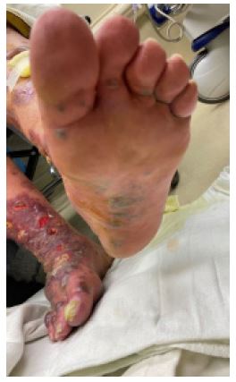
Figure 1: Palpable purpura on soles of feet.
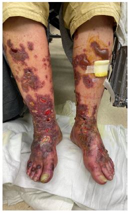
Figure 2: Vesicles and tense bullae extending proximally to the thighs.
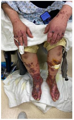
Figure 3: Palpable purpura on dorsum of the hands.
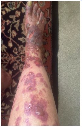
Figure 4: Palpable purpura and tense bullae.
Intralesional and perilesional punch biopsies for histopathology and direct immunofluorescence staining was done. The pathology report showed a subepidermal blister/cleft containing numerous degenerating cells and debris, eosinophils and neutrophils. Interstitial hemorrhage/red blood cell extravasation was noted along with edema of the papillary dermis. A superficial and deep mixed perivascular inflammatory cell infiltrate was present within the dermis. The small vessels exhibited features of leukocytoclastic vasculitis including fibrinoid necrosis of blood vessel walls, endothelial swelling, karyorrhexis of WBC’s and thrombi. Ischemic changes including epidermal necrosis were noted along with collections of inflammatory cells within the epidermis. The overall findings are consistent with bullous LCV. Patient was started on oral prednisone (1 mg/kg) daily with a tapering over 30 days, as well as clobetasol propionate 0.05% cream. Dermatology was consulted and agreed with the treatment. At the two-week follow up, visible improvement of skin findings was noted, and patient’s overall symptoms were resolving with systemic steroids.
Table 1: Laboratory results on admission
| Pertinent Laboratory Markers | Value |
|---|---|
| WBC (103/𝜇L) | 13.1 |
| RBC (106/𝜇L) | 5.51 |
| Hgb (g/dL) | 15 |
| Hct (L/L) | 46.7 |
| Plt Count (103/𝜇L) | 321 |
| Neut % | 58.6 |
| Mono % | 11.4 |
| ESR (mm/hr) | 34 |
| Sodium (mmol/L) | 135 |
| Potassium (mmol/L) | 3.7 |
| Chloride (mmol/L) | 102 |
| Carbon dioxide (mmol/L) | 31 |
| Anion Gap (mEq/L) | 6 |
| BUN (mg/dL) | 23 |
| Creatinine (mg/dl) | 0.97 |
| Glucose (mg/dL) | 113 |
| Hemoglobin A1c % | 5.9 |
| Lactic acid (mmol/L) | 1.4 |
| Calcium (mg/dl) | 9 |
| Total Bilirubin (mg/dl) | 1.6 |
| AST (IU/L) | 40 |
| ALT (IU/L) | 38 |
| Alkaline Phosphatase (IU/L) | 119 |
| C-Reactive Protein | 5.53 |
| Total Protein (g/dL) | 7.7 |
| Albumin (g/dl) | 3.6 |
| Procalcitonin (ng/ml) | < 0.05 |
| Complement C3 (mg/dl) | 142 |
| Complement C4 (mg/dl) | 25.1 |
| Hepatitis A IgM Ab | Non-reactive |
| Hep Bs Antigen | Non-reactive |
| Hep B Core IgM Ab | Non-reactive |
| Hepatitis C Antibody | Non-reactive |
| HIV 1&2 Ag/Ab, 4th Gen | Non-reactive |
| BMI (kg/m2) | 29.2 |
Discussion
Leukocytoclastic vasculitis, also termed hypersensitivity vasculitis, is a small-vessel vasculitis. It involves arterioles, capillaries and postcapillary venules with the inflammatory infiltrate composed of neutrophils with fibrinoid necrosis and disintegration of nuclei into fragments [1,2]. The incidence of LCV ranges from 15 to 38 cases per million/year whereas prevalence 2.7 to 29.7 per million/year [2,3]. In adults it may be associated with systemic necrotizing vasculitis, connective tissue disease, systemic bacterial infection, or malignancy [4]. As mentioned earlier leukocytoclastic vasculitis can be single organ cutaneous in 50% cases with other half associated with systemic involvement. Single organ cutaneous vasculitis can be idiopathic or induced by drugs or infection [5-7].
Skin is the organ most commonly involved in LCV, with typical presentation as a painful, burning rash predominantly in the lower extremities, with up to one-third of patients presenting with trunk and upper extremity involvement [8]. The most common skin manifestation of LCV is palpable purpura, Other skin manifestations include maculopapular rash, bullae, papules, plaques, nodules, ulcers, and livedo reticularis [8]. Our case is a rare manifestation of LCV which presented as tense bullae.
Leukocytoclastic vasculitis needs a detailed clinical evaluation along with skin biopsy for its definitive diagnosis. We need to differentiate limited versus systemic involvement, as well as any causative agent involving the rash which include, ingestion of drugs, infection or any comorbid conditions [2]. The medical history includes symptoms and signs of systemic vasculitis, such as fever, weight loss, and other constitutional symptoms; arthralgia or arthritis; myalgia; abdominal pain, melena or hematochezia; cough, hemoptysis, or dyspnea; hematuria; sinusitis or rhinitis; and paresthesia, weakness or foot drop [2]. If one or more of these symptoms are present, a targeted workup should be performed, in order to identify severe extracutaneous manifestations of systemic vasculitis [2]. Immediate evaluation is required as delay in diagnosis and treatment is associated with significant morbidity and mortality [9]. Lab evaluation includes HBV,HCV, HIV serology, antistreptolysin-O antibodies, fungal serologies, complete blood count, creatinine, urinalysis, sedimentation rate, chest X-ray, liver function test, cutaneous biopsy and direct immunofluorescence test [9]. Further laboratory investigation should be done in combination with the history and physical examination obtained from the patient.
Bullous LV is one of the rare presentations of LV, which can easily be overlooked. It is vital to differentiate it from Bullous pemphigoid which has a similar clinical presentation, via skin biopsy. It would also reveal the size of vessels involvement, immunoglobulin deposit and neutrophil deposit [10].
Treatment of LCV is based on severity of the condition, limited versus systemic involvement, acute versus chronic and the type of skin lesion. LCV has excellent prognosis if the lesion is single, episodic and self-limited [9]. Bed rest, warming, elevation of lower extremities, and NSAIDs analgesic are used to control symptoms, while antihistamine controls pruritis [9].
With significant symptoms, nodular, ulcerative or vesicobullous form of lesions, systemic medication is required. These treatment options require re-evaluation at six to 12 months. The medications that can be used for such conditions are colchicine 0.6-1.8mg/day with expected complete resolution of LCV within 1-2 weeks [11-13]. Dapsone is another option with a few case studies showing improvement in cutaneous LCV with a dose of 100-200 mg daily [9,14,15]. Prednisone is the most widely used treatment with an initial dose of 0.5-1 mg/kg daily with prolonged taper to prevent rebound [9]. It is effective in treating acute or single episode of LCV, however it is not recommended for recurrent or chronic LCV due to its adverse effects [9]. Options of recurrent chronic symptomatic LCV are steroid sparing agents like azathioprine, mycophenolate mofetil and cyclosporine [9].
Limited LCV has a good prognosis if presenting as palpable purpura however, further studies are required to identify the prognosis and outcome of patients with bullous LCV manifestation, and especially identify and address the underlying condition such as malignancy or connective tissue disease that may manifest as LCV initially.
Conclusion
Leukocytoclastic vasculitis should be considered as one of the differential diagnosis of tense bullous skin rash. Skin biopsy is helpful to differentiate the type of lesion with each one having its own treatment course. Our case was unique as it had one of the rare clinical presentations of LV - bullous lesions. We were able to diagnose it only after a skin biopsy. Our patient was treated with systemic steroids with improvement of symptoms in two weeks. Further research is needed in this field, with special regards to the definitive management and prognosis of idiopathic bullous LCV.
Disclosure
Author contributions: All authors wrote, revised, and approved the manuscript, and acquired the data. RO Florica is the article guarantor.
Financial disclosure: None to report.
Informed consent from patient was obtained for this case report.
Conflict of interest: None.
References
- Caproni M, Verdelli A. An update on the nomenclature for cutaneous vasculitis. Curr Opin Rheumatol. 2019; 31: 46-52.
- Fraticelli P, Benfaremo D, Gabrielli A. Diagnosis and management of leukocytoclastic vasculitis. Intern Emerg Med. 2021; 16: 831-841.
- García-Porrúa C, González-Gay MA. Comparative clinical and epidemiological study of hypersensitivity vasculitis versus Henoch-Schönlein purpura in adults. Semin Arthritis Rheum. 1999; 28: 404-412.
- Blanco R, Martínez-Taboada VM, Rodríguez-Valverde V, García-Fuentes M. Cutaneous vasculitis in children and adults. Associated diseases and etiologic factors in 303 patients: Associated diseases and etiologic factors in 303 patients. Medicine (Baltimore). 1998; 77: 403-418.
- Sais G, Vidaller A, Jucglà A, Servitje O, Condom E, Peyri J. Prognostic factors in leukocytoclastic vasculitis: a clinicopathologic study of 160 patients. Arch Dermatol. 1998; 134: 309-315.
- Loricera J, Blanco R, Ortiz-Sanjuán F, Hernández JL, Pina Tet al. Single-organ cutaneous small-vessel vasculitis according to the 2012 revised International Chapel Hill Consensus Conference Nomenclature of Vasculitides: a study of 60 patients from a series of 766 cutaneous vasculitis cases. Rheumatology (Oxford). 2015; 54: 77-82.
- Pastuszczak M, Celińska-Löwenhoff M, Sułowicz J, Wojas-Pelc A, Musiał J. Clinical study on single-organ cutaneous small vessels vasculitis (SoCSVV). Medicine (Baltimore). 2017; 96: e6376.
- Einhorn J, Levis JT. Dermatologic diagnosis: Leukocytoclastic vasculitis. Perm J. Summer. 2015; 19: 77-78.
- Russell JP, Gibson LE. Primary cutaneous small vessel vasculitis: approach to diagnosis and treatment. Int J Dermatol. 2006; 45: 3-13.
- Gota CE, Calabrese LH. Calabrese LH: Diagnosis and treatment of cutaneous leukocytoclastic vasculitis. Int J Clin Rheumtol.
- Sais G. Colchicine in the treatment of cutaneous leukocytoclastic vasculitis: Results of a prospective, randomized controlled trial. Arch Dermatol. 1995; 131: 1399.
- Asherson RA, Buchanan N, Kenwright S, Fletcher CM, Hughes GR. The normocomplementemic urticarial vasculitis syndrome--report of a case and response to colchicine. Clin Exp Dermatol. 1991; 16: 424-427.
- Callen JP. Colchicine is effective in controlling chronic cutaneous leukocytoclastic vasculitis. J Am Acad Dermatol. 1985; 13: 193-200.
- Ruiz Villaverde R, Blasco Melguizo J, Martin Sanchez MC, Naranjo Sintes R. Annular leucocytoclastic vasculitis: response to dapsone. J Eur Acad Dermatol Venereol. 2002; 16: 544-546.
- Fredenberg MF, Malkinson FD. Sulfone therapy in the treatment of leukocytoclastic vasculitis. Report of three cases. J Am Acad Dermatol. 1987; 16: 772-778.
