
Open Access, Volume 9
A peritoneal hydatid cyst mimicking a peritoneal carcinomatosis: Case report and literature review
Ribal Aby Hadeer
University of Balamand Faculty of Medicine and Medical Sciences, Lebanon.
Email: ribal.abyhadeer@std.balamand.edu.lb
Received : April 29, 2023,
Accepted : June 21, 2023
Published : June 30, 2023,
Archived : www.jclinmedcasereports.com
Abstract
Hydatid disease is a zoonoses caused by the larval stage of the cestode Echinococcus. Human become accidental intermediate hosts through the ingestion of foods contaminated by dog faeces containing Echinococcus granulosus eggs or due to close contact with pets carrying the parasite. The cysts of Echinococcus granulosus can be seen in every organ but primary peritoneal hydatid cyst is very rare accounting for only 2% of all intra-abdominal hydatid disease.
Herein, we present the case of a 59-year-old male patient presenting with a suspicious metastatic abdominal mass two month post urgent Hartmann procedure for obstructive sigmoid mass found to have rare primary peritoneal hydatid cyst.
Hydatid disease should be included in the differential diagnosis of peritoneal, mesenteric and omental cyst in endemic countries like the Mediterranean countries, Middle East, Southern part of South America, Iceland, Australia and Africa. The gold standard for final diagnosis is histopathology. The best option for curative treatment is complete resection of all of the lesions in the same setting, but it is not always possible.
Keywords: Echinococcus granulosus; Hydatid cyst; Primary peritoneal hydatid cyst; Surgery; Radical treatment.
Copy right Statement: Content published in the journal follows Creative Commons Attribution License (http://creativecommons.org/licenses/by/4.0). © Ribal AH (2023)
Journal: Open Journal of Clinical and Medical Case Reports is an international, open access, peer reviewed Journal mainly focused exclusively on the medical and clinical case reports.
Citation: Ribal AH. A peritoneal hydatid cyst mimicking a peritoneal carcinomatosis: Case report and literature review. Open J Clin Med Case Rep. 2023; 2060.
Introduction
Hydatid disease is a zoonoses caused by the larval stage of the cestode Echinococcus. The species known to cause infection in humans are Echinococcus granulosus, Echinococcus multilocularis, Echinococcus vogeli and Echinococcus oligarthus [1]. The increase in world travel and migration of people across continents has made hydatid disease endemic in both developed and developing countries [2]. Human become accidental hosts through the ingestion of foods contaminated by dog faeces containing Echinococcus granulosus eggs or due to close contact with pets carrying the parasite [1]. The cysts of Echinococcus granulosus can be seen in every organ but primary peritoneal hydatid cyst is very rare accounting for only 2% of all intra-abdominal hydatid disease [3]. Herein, we present the case of a 59-year-old male patient presenting with a suspicious pelvic mass found to have rare primary peritoneal hydatid cyst.
Case Presentation
59-year-old male patient with history of coronary artery disease, hypertension, dyslipidemia who underwent an urgent Hartmann procedure for an obstructive sigmoid mass one months prior to presentation was found to have 6.6 by 6 by 6.8 cm necrotic mass anterior to the rectum invading the small bowel loops on post operative Ct scan for disease staging. A Positron Emission Tomography (PET) scan was done for better assessment of the mass and showed a large radiotracer in this mass with SUV max 14, probably metastatic. No other metastatic lesions were seen.
Patient reported no episodes of fever, chills, nausea or vomiting. Patient’s vitals were all stable. Physical exam showed soft abdomen, mildly tender and non-distended. Laboratory results showed a white count of 6700 uL with neutrophils 70%, lymphocytes 20%, and eosinophils 3.1%. C-reactive protein was 8 mg/L, Carcinoembryonic antigen was 2.28, CA 19.9 was 11.8 and the rest were unremarkable. No hydatid serology was done.
Colonoscopy from anus until the rectal stump at 25 cm was normal with no recurrence found. Colonoscopy from the colostomy until the cecum was also unremarkable.
A decision was made to go for resection of the mass. Inspection of the abdomen showed extensive small bowel adhesions to the peritoneum with no intra-abdominal collections or ascites or deposits that may suggest recurrence. A pelvic mass in Douglas pouch was identified adherent to the small bowels and rectal stump. Resection of the mass was performed along with enterectomy (Figure 1).
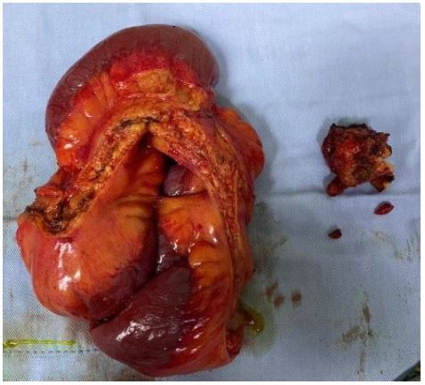
Figure 1: Specimen resected (Mass in the douglas with the adherent small bowel).
Pathology showed inflammatory pseudotumor in the Douglas with few parasitic structures most consistent with echinococcus granulosus scolices and no sign of malignancy. Patient had an uncomplicated hospital stay. He was discharged home on day 5 postoperatively on albendazole treatment for one month then follow up.
Discussion
The major primary site for Echinococcus granulosus in adults is the liver (65-70%) and the secondary site are the lungs (15%) [1]. The cysts of Echinococcus granulosus can also be seen in unusual sites such as peritoneum, omentum, and mesentery of the bowel. Peritoneal hydatidosis accounts for 10-16% of intra-abdominal hydatid disease. Multiples hypothesis has been advocated for the cause of peritoneal hydatidosis. The first one is spontaneous or intra-operative spillage of other cysts this occurs more frequently and is known as secondary peritoneal hydatidosis [4]. The second is through the arterial circulation or via lymphatics and this is known as primary peritoneal hydatidosis which is an extremely rare entity even in endemic areas accounting for just 2% of all intra-abdominal hydatid diseases [3]. Its low prevalence could be explained by the physical barriers to the hematogenous diffusion of cysts created by the liver and lungs [5]. In our case, contamination may be explained by intraoperative spillage of the cysts during his previous surgery for urgent Hartmann procedure due to obstructive colon cancer.
Peritoneal hydatidosis is classified into four groups: localized hydatid cyst, disseminated when the peritoneal cavity is filled with multiple cysts occupying all abdominal quadrants, hydatid carcinomatosis characterized by millimetric cystic lesions disseminated on all surfaces of the peritoneal serosa and hydatidoperitoneum when there is a collection surrounded by an inflammatory membrane, isolating it from the surrounding structures [6]. In our case it was a hydatidoperitoneum and our patient had an inflammatory mass in the Douglas pouch, not disseminated.
The diagnosis of hydatid cyst must be considered especially in endemic regions with a history of raising pets, whenever a cystic mass is felt in the abdominal cavity. The differential diagnosis of such cystic intra-abdominal masses includes pancreatic cyst, mesenteric cyst, gastrointestinal duplication cyst, ovarian cyst, lymphangioma, intra-abdominal abscess, loculated ascites, and hematoma [7].
Clinical presentation of hydatid disease can be non-specific and depends on the cyst size, the site of involvement and the effect on adjacent organs [8,9]. The most common symptom of peritoneal HD is abdominal pain or acute allergic reaction in case of cysts rupture secondary to antigenic fluid that is released in the abdomen [10]. Our patient was asymptomatic and had an incidental finding of necrotic mass on staging PET scan.
The sensitivity of serologic tests is inversely related to the degree of sequestration of the echinococcal antigens inside cysts, regardless of location. Therefore, healthy, intact cysts can elicit a minimally detectable response, whereas previously ruptured or leaking cysts are associated with strong responses. The indirect hemagglutination test is sensitive for the detection of hydatid cyst but has now been replaced by the enzyme immunoassay (ELISA) for initial screening which is more specific. And the hemogram may show evidence of eosinophilia [1].
Ultrasound has a high sensitivity reaching 100%, it can be used in first intention to helps confirm the diagnosis and assess the number, location and anatomic relationship of the cysts [3]. However, the topographic reliability of the ultrasound is lower than that of CT examination that also allows for an optimal analysis of the calcifications and the relationship with the urinary or vascular tract avoiding examinations with more exposure to radiation. On CT, their appearance varies: They may show a “spoke wheel” pattern or a water lily sign. When cysts are healed or in an inactive state, they appear as multiple cystic lesions or with calcification in the peritoneum [11]. During ultrasound evaluation the presence of daughter cysts or the presence of a high and low attenuation rim at the cyst’s periphery is highly indicative of hydatid disease [12].
Despite all these modalities, many differential diagnoses exist specially in cases with complicated cysts. Therefore, the gold standard for final diagnosis is histopathology in which typical cysts with scolices and hooklets are diagnostic [11]. When hydatid disease is the differential diagnosis, biopsy or fine-needle aspiration are not recommended due to the risk of spillage and dissemination of the daughter cysts that can cause anaphylactic reaction and increase recurrence rate [13]. In our patient, the diagnosis was made in the post operative histopathology result for a suspicious metastatic mass of colon cancer.
According to the guidelines of the World Health Organization (WHO), peritoneal localization of hydatid disease is an indication for surgical treatment. Total cystectomy, whenever possible without organ sacrifice is the treatment of choice. Drainage and wide deroofing is recommended when cysts are attached to intraperitoneal viscera or major vessels, or in case of deep localization. Removal of all the cysts during the same intervention is preferred. Otherwise, planned reintervention should be considered [6]. Our patient had a complete cystectomy along with enterectomy due to its proximity to the mass. WHO guidelines also recommend chemotherapy for peritoneal cysts. Preoperative use of albendazole or mebendazole reduce the risk of cystic echinococcosis recurrence and it softens and reduces intracity pressure, thereby simplifying cyst removal. Postoperative chemotherapy is recommended in cases in which spillage may have occurred with albendazole for at least 1 month [14]. Brunetti et al. recommend a long-term use of Albendazole for an indefinite length of time in case of disseminated disease [15]. After diagnosis of Hydatid disease, long term follow up by ultrasound at shorter intervals and CT or MRI at intervals of 2-3 years should be planed [15]. As the diagnosis was made post-operatively, our patient was discharged on albendazole treatment and follow up after one month.
Conclusion
Hydatid disease should be included in the differential diagnosis of peritoneal, mesenteric, and omental cyst in endemic countries. Despise all the imaging and biological tests, the gold standard for final diagnosis is histopathology in which typical cysts with scolices and hooklets are diagnostic. The best option for curative treatment is complete resection of all of the lesions in the same setting, but it is not always possible. The surgeon must adapt the procedures according to the type and localization of the lesions and the patient.
References
- Pedro Moro, Peter M. Schantz, Echinococcosis: a review, International Journal of Infectious Diseases. 2009; 13: 125-133.
- Nahla Kechiche, Dorsaf Makhlouf, Rachida Lamiri, Arije Zouaoui, Lassaad Sahnoun, et al, Peritoneal Hydatid Cysts in Children: A Case Series of Rare Echinococcosis Localization, Iran J Med Sci. 2021; 46.
- H. Benhamichea, D. Sottier, M. Funes De La Vega, B. Cuisenier C, N. Mejean A, et al, Peritoneal hydatidosis and hepatic hydatid cyst perforation, Diagnostic and Interventional Imaging. 2013; 94: 1157-1160.
- Sachar S, Goyal S, Goyal S, Sangwan S. Uncommon Locations and Presentations of Hydatid Cyst, Annals of Medical and Health Sciences Research. 2014; 4.
- Mohammed Anass Majbar, Amine Souadka, Farid Sabbah, Mohamed Raiss, Abdelmalek Hrora, et al , Peritoneal Echinococcosis: Anatomoclinical Features and Surgical Treatment, , World J Surg. 2012; 36: 1030-1035.
- hegde N, Hiremathet B. Primary peritoneal hydatidosis, BMJ Case Rep 2013.
- Murvet Yuksel, Gulen Demirpolat, Ahmet Sever, Sevgi Bakaris, Ertan Bulbuloglu, et al, Hydatid Disease Involving Some Rare Locations in the Body: A Pictorial Essay, Korean J Radiol. 2007; 8.
- Mohamed Ben Khalifa, Mossaab Ghannouchi, Karim Nacef, Aymen Saidi, et al, Small bowel volvulus as a rare manifestation of a primary peritoneal hydatid cyst.
- Bita Geramizadeh, MD, Isolated Peritoneal, Mesenteric, and Omental Hydatid Cyst: A Clinicopathologic Narrative Review, Iran J Med Sci November. 2017; 42.
- Sarthak Sharma, Khalid Mehmood. Fistulization of Peritoneal Hydatid Cyst to the Gastrointestinal Tract: An Unusual Cause of Subacute Intestinal Obstruction, Cureus. 11: e5978
- C. Efthimiadis , G. Anthimidis , K. Vasileiadou, K. Vasileiadou, BG. Koimtzis, et al Management of peritoneal hydatid cysts: A forty-year experience, Heliyon. 2018; 4: e00994.
- Pappas AG, Dede K, Kallimani A, Psaroudaki Z, Argyropoulou A, et al. A 27-Year-Old Man with Intermittent Chest Pain during the Last 4 Years. Chest. 2021; 159: e209-e214.
- Acute rupture of a peritoneal hydatid cyst, Toufik Berri. Department of Surgery, Tourabi Boudjemaa Hospital, Bechar, Algeria. 2014.
- Bashar Almasri , Lina Albitar. Rare isolated primary peritoneal hydatid cysts: A case report from Syria, Qatar Medical Journal. 2016; 2016.
- Brunetti E, Kern P, Vuitton DA. Writing Panel for the WHO-IWGE. Expert consensus for the diagnosis and treatment of cystic and alveolar echinococcosis in humans. Acta Trop. 2010; 114: 1-16.
