
Open Access, Volume 9
A case of mucous membrane pemphigoid masquerading as an adverse food reaction in the allergy clinic
Anita Arthur1; Sarah Fitzpatrick2; Marjorie Montanez-Wiscovich1; Vanessa Cavero-Chavez3; Lyda Cuervo-Pardo3*
1Department of Dermatology, University of Florida College of Medicine, Gainesville, Florida, United States of America.
2College of Dentistry, University of Florida, Gainesville, Florida, United States of America.
3Division of Rheumatology, Allergy & Clinical Immunology, University of Florida College of Medicine, Gainesville, Florida, United States of America.
Lyda Cuervo-Pardo
Division of Rheumatology, Allergy & Clinical Immunology, University of Florida College of Medicine, Gainesville, Florida, United States of America.
Email: lydacp@yahoo.com
Received : April 26, 2023,
Accepted : June 15, 2023
Published : June 20, 2023,
Archived : www.jclinmedcasereports.com
Abstract
Adverse food reactions are a common reason for allergist consultation. Many patients assume their symptoms to be allergic in nature, but severe conditions like mucous membrane pemphigoid (MMP) can be missed if not considered. We report the case of a patient presenting with concern for food allergy found to have MMP.
Keywords: Mucous membrane pemphigoid; Immunobullous disease; Mucosal eruption; Desquamative gingivitis.
Abbreviations: MMP: Mucous Membrane Pemphigoid; BMZ: Basement Membrane Zone; OLCR: Oral Lichenoid Contact Reaction; OLDR: Oral Lichenoid Drug Reaction; PV: Pemphigus Vulgaris; BMS: Burning Mouth Syndrome; OAS: Oral Allergy Syndrome; DIF: Direct Immunofluorescence; IVIG: Intravenous Immunoglobulin.
Copy right Statement: Content published in the journal follows Creative Commons Attribution License (http://creativecommons.org/licenses/by/4.0). © Cuervo-Pardo L (2023)
Journal: Open Journal of Clinical and Medical Case Reports is an international, open access, peer reviewed Journal mainly focused exclusively on the medical and clinical case reports.
Citation: Arthur A, Fitzpatrick S, Montanez-Wiscovich M, Cavero-Chavez V, Cuervo-Pardo L. A case of mucous membrane pemphigoid masquerading as an adverse food reaction in the allergy clinic. Open J Clin Med Case Rep. 2023; 2057.
Introduction
Adverse food reactions are a common reason for allergist consultation. Many patients assume that symptoms involving the oral cavity are related to allergies, but severe conditions like Mucous Membrane Pemphigoid (MMP), a chronic immunobullous condition, can be missed. MMP, also known as cicatricial pemphigoid, is a rare autoimmune blistering disorder involving autoreactive antibodies directed against various autoantigens in the basement membrane zone (BMZ) of skin and mucosae [1,2]. It usually affects elderly patients between the ages of 60 and 80, with a female predominance [1]. The most common site of involvement is the oral mucosa (80-90%), followed by the ocular mucosa (50%), the skin (20%), the genital mucosa (15%), the anal mucosa (10%), and the pharynx, esophagus, and larynx (< 10%) [3]. In the oral cavity, the most commonly affected site is the gingiva (70% of oral MMP cases), then the buccal mucosa (60%), the palate (27%), and the tongue and lips (13%) [4]. Clinical presentation varies, but the oral cavity can have desquamative gingivitis (erythema and erosions of the gingiva) and rarely vesicles and bullae [1]. We report the case of a patient presenting to the allergy clinic with concerns for food allergy found to have MMP.
Case Presentation
A 58-year-old woman presented to the allergy clinic with an 18-month history of intermittent painful vesicles in her mouth, which she associated with food ingestion. Symptoms started when she took up gardening and began eating more fresh vegetables like tomatoes from her garden. She denied rhinitis, hives, and gastrointestinal symptoms. Medications she was taking included apixaban, atorvastatin, levothyroxine, metoprolol succinate, and trospium. Physical exam showed desquamative gingivitis involving the maxillary and mandibular gingiva (Figure 1). Ocular and skin exams were unremarkable. Diagnostic labs, including complete blood count, ferritin, anti-Ro, anti-La, vitamin B12, and folate were unrevealing. Skin testing to inhalants to evaluate cross-reactivity with oral allergens was uninterpretable due to dermatographism. In vitro respiratory allergy panel showed allergic sensitization to trees, ragweed, and grasses. Patch testing was not performed. Given her presenting symptoms and lack of rhinitis, she was referred to the Oral Medicine clinic. On further questioning, she also reported exacerbation of the rash when consuming cinnamon, cucumbers, almonds, and using mint flavored toothpaste. The differential diagnosis included Oral Lichenoid Contact Reaction (OLCR), Oral Lichenoid Drug Reaction (OLDR), irritant contact dermatitis, and autoimmune blistering disorders such as MMP or Pemphigus Vulgaris (PV). Burning Mouth Syndrome (BMS) was considered, but since BMS presents with normal appearing oral mucosa, it was considered unlikely. Oral Allergy Syndrome (OAS) was also considered, but the patient’s symptoms persisted and did not resolve with avoidance of the suspected foods. The physical exam findings also made this condition unlikely. The final diagnosis of mucous membrane pemphigoid (MMP) was made upon gingival biopsy for routine histopathology and direct immunofluorescence. She was started on clobetasol gel and referred to dermatology and ophthalmology for further management.
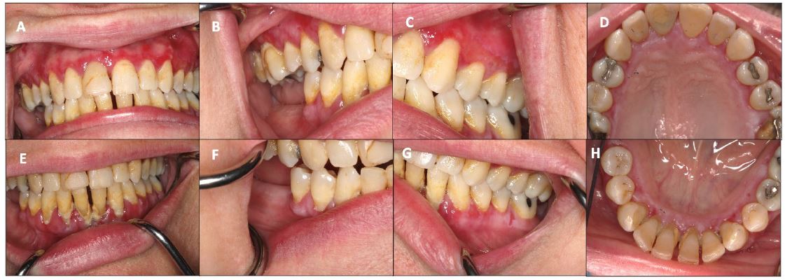
Figure 1: Maxillary and mandibular gingiva with erythema and erosions (Figure 1 A-H).
Discussion
The oral cavity may present with a wide spectrum of conditions. Hypersensitivity or irritant reactions along with autoimmune blistering diseases need to be considered, particularly if a patient presents with desquamative gingivitis. MMP in the oral cavity displays a preference for the gingiva, in many cases presenting only in this area as desquamative gingivitis as in the case of our patient. Biopsy, including Direct Immunofluorescence (DIF) testing, remains the gold standard for diagnosis of MMP demonstrating approximately 60-90% sensitivity [5].
First line treatment options for mild to moderate MMP include dapsone in conjunction with potent topical or intralesional corticosteroids, or tetracyclines plus nicotinamide for mild disease. Short courses of oral steroids may also help [1]. When there is ocular involvement, cyclophosphamide plus systemic steroids or steroid-sparing immunosuppressive medications like mycophenolate mofetil or azathioprine are preferred. Intravenous immunoglobulin (IVIG) can also be used for severe disease [1]. Oral lesions of MMP may lead to scarring, and ocular lesions, if left untreated, may lead to scarring with vision loss [5]. Airway obstruction or esophageal strictures are also possible rare complications [5]. A multidisciplinary approach to care is often necessary for successful treatment outcomes.
This case highlights the need to consider MMP in the differential diagnosis for patients with suspected food allergies involving the oral cavity presenting with desquamative gingivitis.
References
- Alikhan A, Hocker TLH. Blistering Diseases. Chaval R, Dandekar MN, Elbuluk N, Eisen DB, Gandhi RK, Griggin JR, Householder AL, Mir A, Shah KN, Sivamani RK, Tollefson MM I, section editors. In: Review of Dermatology, Elsevier. 2017; 91-92.
- Alrashdan MS, Kamaguchi M. Management of mucous membrane pemphigoid: a literature review and update. Eur J Dermatol. 2022; 32: 312-321.
- Amber KT, Murrell DF, Schmidt E, Joly P, Borradori L. Autoimmune subepidermal bullous diseases of the skin and mucosae: clinical features, diagnosis, and management. Clin Rev Allergy Immunol. 2017; 381: 320-326.
- Hayakawa T, FurumuraM, Fukano H, Li X, Ishii N, et al. Diagnosis of oral mucous membrane pemphigoid by means of combined serologic testing. Oral Surg Oral Med Oral Pathol Oral Radiol. 2014; 117: 483-496.
- Du G, Patzelt S, van Beek N et al. Mucous membrane pemphigoid. Autoimmunity Reviews. 2022; 21: 103036.
