
Open Access, Volume 9
Right atrial diverticulum: A case report and review of literature
Ajmer Singh1*; Mouyeen A Khan2; Manisha Mishra3
1Department of Cardiac Anaesthesia, Medanta-The Medicity, Gurugram (Haryana)-122001, India.
2Institute of Critical Care and Anesthesiology, Medanta-The Medicity, Gurugram (Haryana)-122001, India.
3Medanta-The Medicity, Gurugram (Haryana)-122001, India.
Ajmer Singh
Director, Cardiovascular Anaesthesia, Medanta-The Medicity, Gurgaon (Haryana)-122001, India.
Phone: +91-9818797549, Fax: +91-124-4834111; Email: ajmersingh@yahoo.com
Received : February 24, 2023,
Accepted : April 17, 2023
Published : April 21, 2023,
Archived : www.jclinmedcasereports.com
Abstract
Right atrial diverticulum is a rare congenital abnormality. The presentation can vary from asymptomatic to hemodynamically significant arrhythmia or right heart compression. The diagnosis is suspected by a large cardiac silhouette on the chest X-ray, and confirmed by echocardiography, computed tomography, or magnetic resonance imaging. Herein, we are reporting such a case that was treated surgically. A brief review of the relevant literature is also discussed.
Keywords: Right atrial diverticulum; Cardiac surgery; Surgical excision; Echocardiography.
Copy right Statement: Content published in the journal follows Creative Commons Attribution License (http://creativecommons.org/licenses/by/4.0). © Singh A (2023)
Journal: Open Journal of Clinical and Medical Case Reports is an international, open access, peer reviewed Journal mainly focused exclusively on the medical and clinical case reports.
Citation: Singh A, Khan MA, Mishra M. Right atrial diverticulum: A case report and review of literature. Open J Clin Med Case Rep. 2023; 2020.
Introduction
Diverticulum of the right atrium (RA) is a rare congenital anomaly with only about 30 cases reported in the literature [1]. Patients with a single diverticulum may remain asymptomatic till late in life, while those with symptoms can present in early infancy or childhood. If left untreated, the RA diverticulum carries the risk of thrombus formation, arrhythmia, hemodynamic derangement, life-threatening rupture, and sudden cardiac death. Regardless of the symptoms, the surgical resection of the large diverticulum is recommended.
Сase Report
A 17-year-old female presented with symptoms of palpitations and dyspnea on exertion (New York Heart Association class III) for three months duration. The electrocardiogram showed sinus tachycardia. Chest X-ray revealed marked cardiomegaly (cardiothoracic ratio 0.7), and a large outpouching of the RA (Figure 1). Two-dimensional transthoracic echocardiography (TTE) showed a diverticulum (10 x 8 x 6 cm), communicating with the free wall of RA. The tricuspid valve was normal. The patient was offered the surgical option of resection of the diverticulum, and her family consented to the same.
Intraoperative cardiac monitoring included transesophageal echocardiography (TEE), which showed a large RA diverticulum with a wide neck and spontaneous echo contrast (Figure 2A, 2B). In addition, it showed compression of the right ventricle (Supplementary video 1). The diverticulum was resected from within the RA and the communication was closed with a pericardial patch under hypothermic cardiopulmonary bypass (CPB). Histopathological examination of the diverticulum showed fibrous connective tissue and intima without a muscular layer. At three months’ follow-up, the patient was asymptomatic and TTE was normal.
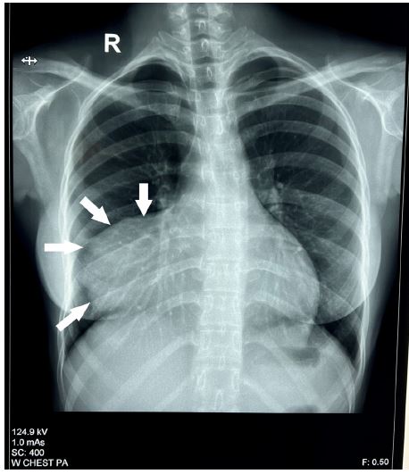
Figure 1: Chest X-ray showing cardiomegaly and outpouching of the right atrium (arrows).
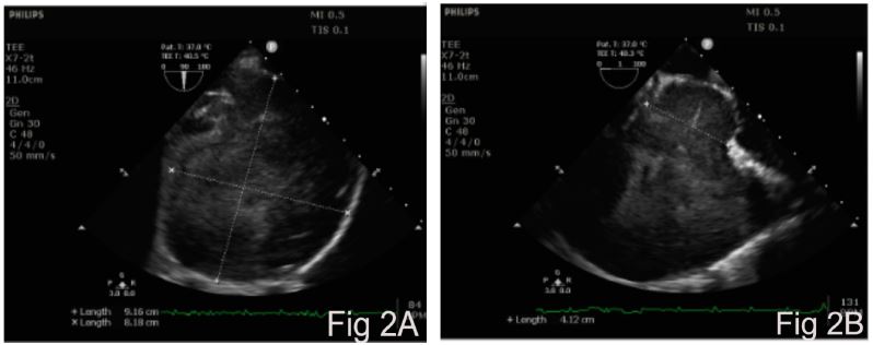
Figure 2: Intraoperative transesophageal echocardiography image showing large right atrial diverticulum (Fig 2A) with wide neck (Fig 2B).
Discussion
Since its first description by Bailey in 1953, only a few cases of RA diverticulum have been reported [2]. The etiology and management of this condition are not clearly defined. The suggested pathogenesis includes an atrial variety of Uhl’s anomaly or persistence of the left Cuvier vein [3]. An absence of myocardial elements in the wall of the diverticulum favors the theory of localized Uhl’s anomaly. Differential diagnosis of RA enlargement includes Ebstein’s anomaly, cor triatriatum dexter, RA aneurysm, pericardial cyst, and mediastinal tumor. Echocardiography can differentiate these anomalies, however, computed tomography, magnetic resonance imaging, or angiography may be required in some instances.
The common arrhythmias associated with RA diverticulum include supraventricular tachycardia, atrial flutter, atrial fibrillation, Wolff-Parkinson-White syndrome, and left bundle branch block. The most plausible explanation for atrial arrhythmias is the presence of an enormous mass of atrial tissue which acts as a substrate for arrhythmias. The arrhythmias generally result from circus movements (atrial re-entrant tachycardias) or by direct stimulation of the cardiac surface. Surgical excision of the diverticulum removes the substrate and generally there is no recurrence of arrhythmias after surgery [4]. Compression of the RA and right ventricle can cause a decrease in ventricular compliance leading to low cardiac output. Sudden cardiac death is reported more frequently in patients with diverticula of the coronary sinus (18%), compared to those with diverticula of the RA (6%) [5].
Most patients with single diverticulum remain asymptomatic till late in life. Of the 105 cases of congenital anomalies of RA and coronary sinus, 13 had single RA diverticula; and among them 84% were symptomatic [5]. Diagnosis is usually incidental during routine cardiac evaluation or chest X-ray [6]. Known complications are mechanical compression of the adjacent chambers, thromboembolism, atrial arrhythmias, and sudden cardiac death. Treatment is surgical excision in symptomatic cases or asymptomatic cases with a huge RA diverticulum. There are reports of resection of the diverticulum without the use of CPB. Total endoscopic removal of the diverticulum on the beating heart is also reported [1]. However, large diverticula and those at risk of rupture should be resected under CPB.
In conclusion, the diverticulum of RA is associated with the risk of thrombosis, atrial arrhythmias, rupture, and sudden cardiac death. Surgical resection of the diverticulum is helpful in symptomatic patients and in patients with a huge diverticulum regardless of symptoms.
References
- Dang HQ, Tran MT, Le HT. Totally endoscopic resection of right atrial diverticulum: A Case Report. Innovations. 2022; 17: 159-161.
- Bailey CP. Surgery of the Heart. Philadelphia, Lea & Febiger. 1955; 413.
- Kodandarama UM, Chaudhury AG, Bhat P, Nanjappa MC. A rare case of the right atrial diverticulum encroaching the left ventricle. J Indian Acad Echocardiogr Cardiovasc Imaging. 2022; 6: 129-131.
- Aggarwal N, Joshi R, Joshi RK, Agarwal M. Right atrial diverticulosis and early-onset arrhythmia: Rare cause of incessant neonatal arrhythmia. Indian Pediatr. 2017; 54: 503-504.
- Binder TM, Rosenhek R, Frank H, Gwechenberger M, Maurer G, Baumgartner H. Congenital malformations of the right atrium and the coronary sinus: an analysis based on 103 cases reported in the literature and two additional cases. Chest. 2000; 117: 1740-1748.
- Honda A, Shojima T, Tuhara N, Morita K, Nakayoshi T, Tahara A, et al. Life-threatening huge right atrial diverticulum. E Heart J Case Reports. 2020; 4: 1-2.
