
Open Access, Volume 9
Long term outcomes of stenting of a single internal carotid artery in complex head and neck paragangliomas
Gianluca Piras1*; Golda Grinblat2; Lorenzo Lauda1; Antonio Caruso1; Dikran Mardighian3; Mario Sanna1
1Department of Otology and Skull Base Surgery, Gruppo Otologico and Mario Sanna Foundation, Piacenza-Rome, Italy.
2Department of Otorhinolaryngology, Hillel Yaffe Medical Center, Hadera, Israel.
3Department of Neuroradiology, Azienda Ospedaliera Spedali Civili di Brescia, Brescia, Italy.
Gianluca Piras
Otologist and Skull Base Surgeon, Gruppo Otologico and Mario Sanna Foundation, Piacenza-Rome, Casa di Cura “Piacenza” S.P.A., Piacenza, Italy.
Tel: +393475846405;
Email: gianlu.piras@libero.it; gianlu.piras@gmail.com
Received : March 08, 2023,
Accepted : April 14, 2023
Published : April 17, 2023,
Archived : www.jclinmedcasereports.com
Abstract
Background: The introduction of preoperative reinforcement of the internal carotid artery (ICA) with stents has significantly improved surgical management of head and neck paragangliomas.
Case report: The aim of this article is to report the management and long term outcomes of a complex case of right ipsilateral tympanojugular and vagal paragangliomas in a patient with absence of the contralateral ICA due to a previous removal of a left carotid body tumor. Surgical removal of the lesions was performed after stenting of the right cervical and intratemporal segment of the ICA. For more than 10 years the patient has not shown disease relapse or complications.
Conclusion: This report demonstrates that even cases considered “inoperable” can be surgically managed with a proper pre-operative plan. This is the first clinical report regarding long term efficacy of endoluminal stents on a single ICA for head and neck paragangliomas.
Keywords: Head and neck paragangliomas; Internal carotid artery; Stenting.
Copy right Statement: Content published in the journal follows Creative Commons Attribution License (http://creativecommons.org/licenses/by/4.0). © Piras G (2023)
Journal: Open Journal of Clinical and Medical Case Reports is an international, open access, peer reviewed Journal mainly focused exclusively on the medical and clinical case reports.
Citation: Piras G, Grinblat G, Lauda L, Caruso A, Mardighian D, Sanna M. Long term outcomes of stenting of a single internal carotid artery in complex head and neck paragangliomas. Open J Clin Med Case Rep. 2023; 2019.
Introduction
Tympanojugular (TJP) and vagal paragangliomas (VP) are benign, slow-growing tumors. Due to their indolent nature, they often present in advanced stages with cranio-temporo-cervical extensions, having the potential to invade surrounding neurovascular structures like the Jugular Bulb (JB), the internal carotid artery (ICA), the facial (FN) and the lower cranial nerves (LCNs) [1].
Nowadays, extensive involvement of the ICA does not represent an absolute contraindication to surgical therapy. TJP and VP frequently involve the ICA due to their close anatomical proximity [2]. When indicated, the tumor must be dissected from the arterial wall. This can be achieved by subperiosteal (or supra-adventitial) dissection of the ICA in the carotid canal (horizontal portion) or subadventitial dissection in the vertical portion [3]. When the artery is completely surrounded by a tumor resulting in severe stenosis on arteriography, manipulation without proper endovascular management may lead to severe bleeding, incomplete removal, or a cerebral vascular accident [4]. Permanent Balloon Occlusion (PBO) is performed when the ICA is infiltrated by the tumor and the collateral blood flow is sufficient. But in cases with insufficient collateral blood flow, we routinely use intraluminal stenting. Stenting of the cervical and petrous segments of ICA was introduced as a preoperative management protocol by the Gruppo Otologico in the clinical and surgical management of complex head and neck paragangliomas since the early 2003 as a way to avoid both preoperative closure of the ICA or high-risk bypass procedures, and to protect and preserve the integrity of the artery during surgery, mainly in cases in which the collateral flow through the circle of Willis is insufficient [5-7]. Stenting of the ICA allows reinforcement of the artery, reducing the risk of intraoperative injury of its wall while performing a more aggressive carotid dissection in the subadventitial plane.
In 2007 we described the case of a patient with a single ICA affected by two ipsilateral paragangliomas (TJP and VP), which were successfully removed without complications after endoluminal stenting of the ICA [6]. For more than 10 years the patient has not shown disease relapse or complications. The aim of this article is to demonstrate long term efficacy of endoluminal stenting of the ICA, making possible what is usually considered “inoperable”.
Case Report
This case was approved from review by the Casa di Cura “Piacenza” S.P.A. Institutional Review Board.
A 40 years-old male patient was referred in 2006 at the Gruppo Otologico, a quaternary referral center for neurotology and skull-base surgery, with a diagnosed right-sided TJP and VP. The patient had been surgically treated elsewhere 15 years before for a left Carotid Body Paraganglioma (CBP) with closure of the left common carotid artery. Clinical examination confirmed impairment of the right LCNs, with compensated vocal cord paralysis.
High-Resolution Computed Tomography (HRCT) of the temporal bone and skull base and gadolinium-enhanced Magnetic-Resonance Imaging (MRI) of the brain and neck showed involvement of the vertical and horizontal portions of the petrous segment of the ICA and two distinct masses at the level of the parapharyngeal space and jugular foramen, suggestive for a TJP (Class C3De1) and VP (Stage I) (Figure 1).
Angiography showed occlusion of the left common carotid artery and ICA. The distal cervical segment and the vertical portion of the petrous segment of the right ICA were irregular and stenotic due to tumor encasement (Figure 1). The patient underwent stenting of the right ICA and received antiplatelet therapy starting 1 week before the procedure. Seven weeks after stenting, the patient underwent embolization of the right ascending pharyngeal and occipital arteries. Two days later he was operated through a right infratemporal fossa type a approach and total removal was achieved with no complications. Right FN function improved from grade VI House-Brackmann scale in the early post-operative period to grade III two years after surgery. The right vocal cord palsy was well tolerated thanks to the contralateral compensation with no need for pharyngolaryngeal surgery. Doppler ultrasound at discharge and at 6, 18 and 36 months after surgery showed a patent right ICA. The patient underwent yearly HRCT and MRI scans for the first 5 years and approximately every 2 years for the subsequent period.
The last contrast-enhanced MRI and HRCT scans (October 2021) demonstrated patency of the right ICA with the intraluminal stenting in place and no signs of tumor recurrence (Figure 2).
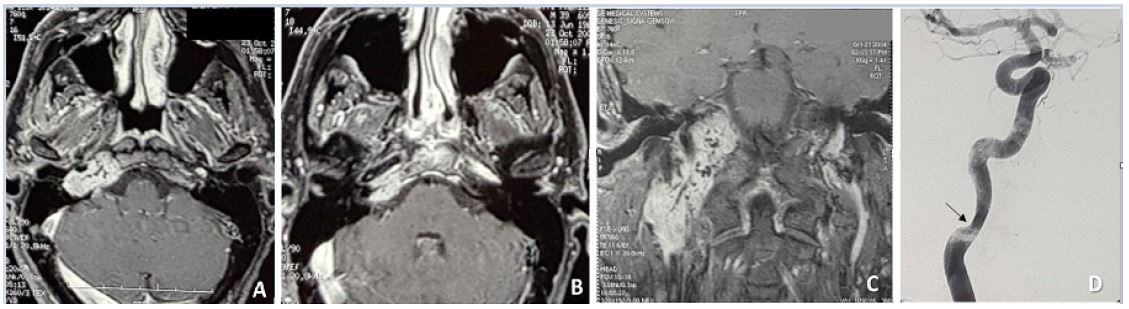
Figure 1: (A,B,C,D): C3De1 TJP (A,B), C3De1 TJP and VP stage I (C) on Gd-enhanced MRI scans. Stenosis of the distal cervical C1 segment of ICA (arrow) can be appreciated on angiography (D).
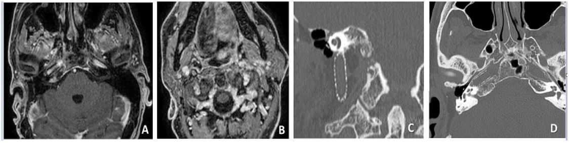
Figure 2: (A,B,C,D): No residual or recurrent tumor can be appreciated on post-operative Gd-enhanced MRI scans (A,B). Post-operative HRCT shows intraluminal stenting from the neck (C) to the horizontal petrous (D) portions of the ICA.
Discussion
This is the first case described in literature regarding long term efficacy of stenting of a single ICA for head and neck paragangliomas. The introduction of preoperative reinforcement of the ICA with stents has significantly improved surgical management of paragangliomas. From a total of 320 TJPs, 31 VPs and 15 CBPs surgically treated in our Institution, 35 cases have been pre-operatively stented with no complications even on a long-term follow-up. Since we first started publishing our experience with endovascular neuroradiological treatment of the ICA in paragangliomas, only a few articles have been published from other authors [10,11]. Cases of class C3-C4 TJPs, Stage II or III VPs or Shamblin class II and III CBPs can be safely removed without risks of intraoperative ICA injury. For decades these classes of tumors have been considered inoperable and many authors suggested partial resection of the tumor followed by radiotherapy or radiotherapic treatment alone [8,9]. A recent meta-analysis done by Shapiro S et al [9]. Regarding outcomes of primary Radiosurgery (RS) for TJPs treatment showed that RS is far from demonstrating any significant advantage in the management of TJPs, both in the short and long term. From 959 selected studies, only 15 met the revised criteria for inclusion in the meta-analysis (with severe lacks on tumor classification, follow-up, tumor volume changes), demonstrating that over 98% of the literature regarding RS for TJPs did not show any scientific and clinical relevance. Moreover, our ongoing studies on the biology of paragangliomas indicate that these tumors express proteins that confer intrinsic radioresistance and drive the epithelial to mesenchymal transition, due to the activation of the NOTCH signaling and ZEB1 expression, which are well known markers of radioresistance [13,14].
Stenting of the ICA is a safe procedure, providing reinforcement to the arterial wall, avoiding the risk of accidental rupture, and offering a limit for the deep resection, making the subadventitial plane of cleavage easier to follow. The presence of endoluminal stent avoids the risk of postoperative delayed aneurism formation or intimal dissection, following subadventitial surgical dissection.
Patients with inadequate contralateral compensatory blood flow represent a further challenge for surgical management: permanent ICA occlusion and en-bloc resection of the artery cannot be performed, therefore tumor resection must be extremely careful to avoid fatal vascular accidents. ICA stenting opens the possibility of treating cases previously considered inoperable, as we demonstrated in this patient.
Endoluminal stenting cannot be performed in any case. The presence of extensive blood supply from branches of the ICA, severe luminal stenosis, massive parietal infiltration, vessel wall weakness because of previous treatment or tortuous course or kinking of the ICA at the skull base represent a contraindication for stenting. PBO of the ICA or partial resection of the tumor represent an alternative.
Disclosure: There is no conflict of interest involved and the present work has not received any funding from any sources.
References
- Sanna M, Piazza P, Shin SH, Flanagan S, Mancini F. Microsurgery of skull base paraganglioma. Stuttgart. Thieme. 2013.
- Sanna M, Jain Y, De Donato G, Rohit LL, Taibah A. Management of jugular paragangliomas: the Gruppo Otologico experience. Otol Neurotol. 2004; 25: 797-804.
- Fisch U, Fagan P, Valavanis A. The infratemporal fossa approach for the lateral skull base. Otolaryngol Clin North Am. 1984; 17: 513-552.
- Sanna M, Piazza P, De Donato G, Menozzi R, Falcioni M. Combined endovascular-surgical management of the internal carotid artery in complex tympanojugular paragangliomas. Skull Base. 2009; 19: 26-42.
- Sanna M, Khrais T, Menozi R, Piaza P. Surgical removal of jugular paragangliomas after stenting of the intratemporal internal carotid artery: a preliminary report. Laryngoscope. 2006; 116: 742-746.
- Piazza P, Di Lella F, Menozzi R, Bacciu A, Sanna M. Absence of the contralateral internal carotid artery: a challenge for management of ipsilateral glomus jugulare and glomus vagale tumors. Laryngoscope. 2007; 117: 1333-1337.
- Konishi M, Piazza P, Shin SH, Sivalingam S, Sanna M. The use of internal carotid artery stenting in management of bilateral carotid body tumors. Eur Arch Otorhinolaryngol. 2011; 268: 1535-1539.
- Prasad SC, Mimoune HA, D’Orazio F, Medina M, Bacciu A, et al. The role of wait-and-scan and the efficacy of radiotherapy in the treatment of temporal bone paragangliomas. Otol Neurotol. 2014; 35: 922-931.
- Shapiro S, Kellermeyer B, Ramadan J, Jones G, Wiseman B, et al. Outcomes of Primary Radiosurgery Treatment of Glomus Jugulare Tumors: Systematic Review With Meta-analysis. Otol Neurotol. 2018; 39: 1079-1087.
- Ong HS, Fan XD, Ji T. Radical resection of a Shamblin type III carotid body tumour without cerebro-neurological deficit: Improved technique with preoperative embolization and carotid stenting. Int J Oral Maxillofac Surg. 2014; 43: 1427-1430.
- McDougall CM, Liu R, Chow M. Covered carotid stents as an adjunct in the surgical treatment of carotid body tumors: a report of 2 cases and a review of the literature.
- Bacciu A, Prasad SC, Sist N, Rossi G, Piazza P, Sanna M. Management of the cervico-petrous internal carotid artery in class C tympanojugular paragangliomas. Head Neck. 2016; 38: 899-905.
- Cama A, Verginelli F, Lotti LV, Napolitano F, Morgano A, et al. Integrative genetic, epigenetic and pathological analysis of paraganglioma reveals complex dysregulation of NOTCH signaling. Acta Neuropathological. 2013; 126: 575-594.
- Verginelli F, Perconti S, Vespa S, Schiavi F, Prasad SC, et al. Paragangliomas arise through an autonomous vasculo-angio-neurogenic program inhibited by imatinib. Acta Neuropathol. 2018; 135: 779-798.
